
Page 31 - Simplicity is Key in CRT
P. 31
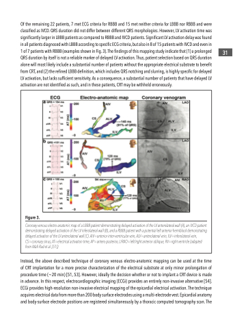
Of the remaining 22 patients, 7 met ECG criteria for RBBB and 15 met neither criteria for LBBB nor RBBB and were classified as IVCD. QRS duration did not differ between different QRS morphologies. However, LV activation time was significantly larger in LBBB patients as compared to RBBB and IVCD patients. Significant LV activation delay was found in all patients diagnosed with LBBB according to specific ECG criteria, but also in 8 of 15 patients with IVCD and even in 1 of 7 patients with RBBB (examples shown in Fig. 3). The findings of this mapping study indicate that (1) a prolonged QRS duration by itself is not a reliable marker of delayed LV activation. Thus, patient selection based on QRS duration alone will most likely include a substantial number of patients without the appropriate electrical substrate to benefit from CRT, and (2) the refined LBBB definition, which includes QRS notching and slurring, is highly specific for delayed LV activation, but lacks sufficient sensitivity. As a consequence, a substantial number of patients that have delayed LV activation are not identified as such, and in these patients, CRT may be withheld erroneously.
Instead, the above described technique of coronary venous electro-anatomic mapping can be used at the time of CRT implantation for a more precise characterization of the electrical substrate at only minor prolongation of procedure time (~20 min) [51, 53]. However, ideally the decision whether or not to implant a CRT device is made in advance. In this respect, electrocardiographic imaging (ECGi) provides an entirely non-invasive alternative [54]. ECGi provides high-resolution non-invasive electrical mapping of the epicardial electrical activation. The technique acquires electrical data from more than 200 body surface electrodes using a multi-electrode vest. Epicardial anatomy and body-surface electrode positions are registered simultaneously by a thoracic computed tomography scan. The
  Figure 3.
Coronary venous electro-anatomic map of a LBBB patient demonstrating delayed activation of the LV anterolateral wall (A), an IVCD patient demonstrating delayed activation of the LV inferolateral wall (B), and a RBBB patient with a potential left anterior hemiblock demonstrating delayed activation of the LV anterolateral wall (C). AIV=anterior inter-ventricular vein, ALV=anterolateral vein, ILV=inferolateral vein, CS=coronary sinus, AT=electrical activation time, AP=antero-posterior, L/RAO=left/right anterior oblique, RV=right ventricle (adapted from Mafi Rad et al. [51])
 31
