
Page 96 - Simplicity is Key in CRT
P. 96
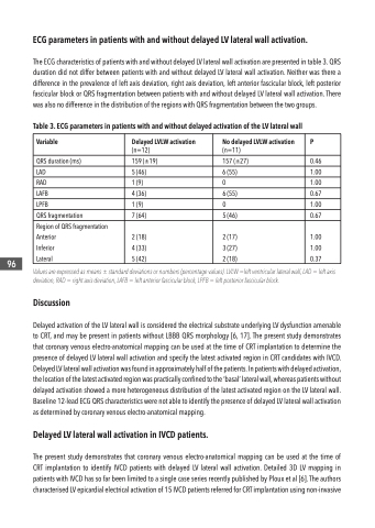
Variable
Delayed LVLW activation
(n=12)
No delayed LVLW activation
(n=11)
P
QRS duration (ms)
159 (±19)
157 (±27)
0.46
LAD
5 (46)
6 (55)
1.00
RAD
1 (9)
0
1.00
LAFB
4 (36)
6 (55)
0.67
LPFB
1 (9)
0
1.00
QRS fragmentation
7 (64)
5 (46)
0.67
Region of QRS fragmentation Anterior
Inferior
Lateral
2 (18) 4 (33) 5 (42)
2 (17) 3 (27) 2 (18)
1.00 1.00 0.37
96
 ECG parameters in patients with and without delayed LV lateral wall activation.
The ECG characteristics of patients with and without delayed LV lateral wall activation are presented in table 3. QRS duration did not differ between patients with and without delayed LV lateral wall activation. Neither was there a difference in the prevalence of left axis deviation, right axis deviation, left anterior fascicular block, left posterior fascicular block or QRS fragmentation between patients with and without delayed LV lateral wall activation. There was also no difference in the distribution of the regions with QRS fragmentation between the two groups.
Table 3. ECG parameters in patients with and without delayed activation of the LV lateral wall
           Values are expressed as means ± standard deviations or numbers (percentage values). LVLW =left ventricular lateral wall, LAD = left axis deviation, RAD = right axis deviation, LAFB = left anterior fascicular block, LPFB = left posterior fascicular block.
Discussion
Delayed activation of the LV lateral wall is considered the electrical substrate underlying LV dysfunction amenable to CRT, and may be present in patients without LBBB QRS morphology [6, 17]. The present study demonstrates that coronary venous electro-anatomical mapping can be used at the time of CRT implantation to determine the presence of delayed LV lateral wall activation and specify the latest activated region in CRT candidates with IVCD. Delayed LV lateral wall activation was found in approximately half of the patients. In patients with delayed activation, the location of the latest activated region was practically confined to the ‘basal’ lateral wall, whereas patients without delayed activation showed a more heterogeneous distribution of the latest activated region on the LV lateral wall. Baseline 12-lead ECG QRS characteristics were not able to identify the presence of delayed LV lateral wall activation as determined by coronary venous electro-anatomical mapping.
Delayed LV lateral wall activation in IVCD patients.
The present study demonstrates that coronary venous electro-anatomical mapping can be used at the time of CRT implantation to identify IVCD patients with delayed LV lateral wall activation. Detailed 3D LV mapping in patients with IVCD has so far been limited to a single case series recently published by Ploux et al [6]. The authors characterised LV epicardial electrical activation of 15 IVCD patients referred for CRT implantation using non-invasive
