
Page 14 - CJO_SP18
P. 14
C CLINICAL RESEARCH
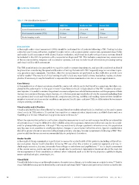
Table 1: TBI Classification System 6,7
Characteristic Mild TBI Moderate TBI Severe TBI
Loss of consciousness (LOC) 0 to 30 mins 0.5 to 24 h >24 h
Post-traumatic amnesia (PTA) 0-1 days 1-7 days >7 days
Brain-imaging results Normal Abnormal Abnormal
EVALUATION
A thorough ocular-visual assessment (OVA) should be performed for all patients following a TBI. Testing includes
a detailed case history, refraction, routine binocular vision and accommodation assessment, automated visual fields
and ocular health assessment with dilated fundus evaluation. Additional in-depth testing of some systems should
be included in the OVA for patients with a suspected or diagnosed TBI. This includes complementary assessments
of the accommodative, vergence and oculomotor systems, and may include visual information-processing testing
and visual-midline shift assessment.
The TBI population is also susceptible to cognitive and/or memory impairments, and special consideration should
be given when considering the speed and duration of testing. Patients with TBI frequently require more time to pro-
cess questions and commands. Therefore, objective measurements are preferred, as they will often provide more
reliable results. The results of clinical testing should include any reported dizziness, headaches, nausea, or photo-
10
phobia. If necessary, it may be beneficial to separate the vision examination into two or more appointments.
Case history
A thorough review of visual symptoms should be conducted, which can be facilitated by a symptom checklist com-
pleted by the patient prior to the appointment. Case history should include details of the TBI incident and associ-
11
ated injuries. It is useful to review the patient’s current and previous rehabilitation services and the progress of their
therapy (occupational therapy, physiotherapy, etc.). Patient goals and needs should also be assessed, including their
occupational and vocational visual demands, computer use, driving, mobility and reading. Optometrists should re-
member to record previous ocular conditions and general health (pre- and post-TBI), to differentiate between new
and pre-existing conditions.
Visual acuity and refraction
Visual acuity itself is less often affected by TBI, and therefore traditional methods (i.e. Snellen) can be used to assess
visual acuity in TBI patients. If a patient has cognitive or communication impairments, modified charts such as a
Tumbling E or Broken Wheel test may provide more valid results.
11
When the optometrist performs a refraction, objective measurements such as retinoscopy should be considered for
all patients since it may be difficult to elicit reliable subjective responses. Automated refractors can also be consid-
ered for photophobic patients. Although TBIs may not directly change a patient’s refractive error, this population
may become more sensitive to small prescription changes or uncorrected refractive errors. Special consideration
should be given to latent or uncorrected hyperopic patients, who may become symptomatic following a TBI. Pro-
12
gressive addition lenses are not recommended due to peripheral distortions.
Ocular health
A thorough slit lamp examination is performed to assess the ocular health of TBI patients, including a dilated fun-
dus evaluation. Ocular health disorders following TBI can affect the anterior or posterior segments and may in-
clude angle recession, dry eye, intraocular hemorrhage or embolisms and papilledema. 13-17 An in-depth assessment
of the cranial nerves, pupils and optic nerves should also be performed. Appropriate treatment should be made for
the management of these conditions or referral when indicated, such as to the family physician, ophthalmologist,
neuro-ophthalmologist, neurologist, etc.
Visual field
Visual field defects may occur through trauma to the optic nerve, chiasm, optic radiations, or occipital cortex. 15-17 Subtle
visual field defects may not be detected by confrontational visual field. Automated perimetry is better suited for de-
18
14 CANADIAN JOURNAL of OPTOMETRY | REVUE CANADIENNE D’OPTOMÉTRIE VOL. 80 NO. 1
37529_CJO_SP18 February 20, 2018 10:55 AM APPROVAL: ___________________ DATE: ___________________