
Page 27 - CJO_SP18
P. 27
CASE REPORT
CASE REPORT
In 1973, a 43-year-old male showed an unusual entrance of the central retinal vessels on his nerve heads (Fig. 1 top
left). Corrected visual acuity for the left eye at that time was 20/15, while that for the right eye was on the order of
20/200. The poor acuity in the right eye may have been related to a car accident at age 4. The right eye also showed
a unilateral esotropia. Both eyes were hyperopic in the 6 D range.
Since the rest of his family were being seen at the University of Waterloo Optometry Clinic at that time, their nerve
heads were also photographed. His wife is shown in Fig. 1 top right. The couple had two daughters and four sons.
All showed some degree of situs inversus of the nerve head blood vessels (Fig. 1 balance of photos). This is consistent
with an autosomal dominant pattern of inheritance. These findings have been reported previously. 5
In 1994, at age 64 y, the patient had a non-secretory (i.e. null cell) pituitary macroadenoma removed via transsphenoidal
resection. The sequence of events leading to discovery of the tumor was as follows. The patient underwent cataract sur-
gery in September 1994. Over the two months following cataract surgery, he experienced a decrease in left visual acuity
from 20/50 to 20/200. Cystoid macular edema was ruled out. Visual field testing for the left eye (Fig. 2) showed decreased
sensitivity in the superior nasal field, close to the center of the field. There was no afferent pupil defect, and extraocular
muscle function was normal. When the patient mentioned to his eye surgeon that his brother had been diagnosed with
a brain tumor in 1990, the eye surgeon ordered a brain scan to rule out a central cause for the loss of acuity. This scan
revealed a macroadenoma, which was removed in late November 1994. Further visual field testing prior to and follow-
ing removal of the macroadenoma (Fig. 3) showed a greatly improved left visual field after surgery; left visual acuity
improved to 20/25, where it remains to date.
The vascular pattern of the optic nerve head is established by the seventh to eighth month of gestation. Although
6
the nerve head anomaly in this patient appears to have been present at birth, the pituitary problem was not noted
until 64 years later.
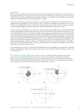
Fig. 3. Goldmann visual field test results closer to the time of surgery: A and B show the right and left field results 3 days
prior to surgery; C is the left field result 3 days after surgery. In A, there is a central defect extending superotemporally.
In B, there is a left superotemporal field defect. In C, the superotemporal field defect has resolved. Shaded areas are scotomas.
Inner solid lines show isopters obtained with the Goldmann II-4 target; outer, broken lines are norms for 65 year olds obtained
with the Goldmann III-3 target, which has a stimulus value equivalent to the II-4 target. 16
15
17
CANADIAN JOURNAL of OPTOMETRY | REVUE CANADIENNE D’OPTOMÉTRIE VOL. 80 NO. 1 27
37529_CJO_SP18 February 20, 2018 10:55 AM APPROVAL: ___________________ DATE: ___________________ Fig. 3. Goldmann visual field test results closer to the time of surgery: A and B
show the right and left field results 3 days prior to surgery; C is the left field
result 3 days after surgery. In A, there is a central defect extending
superotemporally. In B, there is a left superotemporal field defect. In C, the
superotemporal field defect has resolved. Shaded areas are scotomas. Inner
solid lines show isopters obtained with the Goldmann II-4 target; outer, broken