
Page 2 - Alga NPS interactions_Neat
P. 2
Bull Environ Contam Toxicol (2015) 94:554–558 555
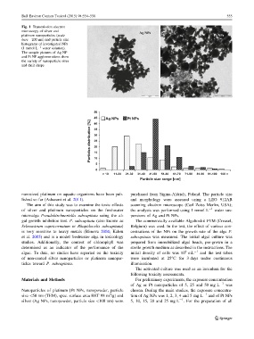
Fig. 1 Transmission electron
microscopy of silver and
platinum nanoparticles (scale Ag NPs Pt NPs
bars—200 nm) and particle size
histograms of investigated NPs
(1 mmol L -1 water solution).
The sample pictures of Ag NP
and Pt NP agglomerations show
the variety of nanoparticle sizes
and their shape
50
45 Ag NPs Pt NPs
Particles distribution [%] 30
40
35
25
20
15
10
5
0
> 10 11-20 21-30 31-40 41-50 51-60 61-70 71-80 81-90 91-100 100 <
Particle size range [nm]
nanosized platinum on aquatic organisms have been pub- purchased from Sigma-Aldrich, Poland. The particle size
lished so far (Asharani et al. 2011). and morphology were assessed using a LEO 912AB
The aim of this study was to examine the toxic effects scanning electron microscope (Carl Zeiss Merlin, USA);
of silver and platinum nanoparticles on the freshwater the analysis was performed using 1 mmol L -1 water sus-
microalga Pseudokirchneriella subcapitata using the al- pensions of Ag and Pt NPs.
gal growth inhibition test. P. subcapitata (also known as The commercially available Algaltoxkit FTM (Creasel,
Selenastrum capricornutum or Rhapidocelis subcapitata) Belgium) was used. In the test, the effect of various con-
is very sensitive to heavy metals (Blinova 2004; Kahru centrations of the NPs on the growth rate of the alga P.
et al. 2005) and is a model freshwater alga in toxicology subcapitata was measured. The initial algal culture was
studies. Additionally, the content of chlorophyll was prepared from immobilized algal beads, pre-grown in a
determined as an indicator of the performance of the sterile growth medium as described in the instructions. The
6
algae. To date, no studies have reported on the toxicity initial density of cells was 10 mL -1 and the test tubes
of non-coated silver nanoparticles or platinum nanopar- were incubated at 25°C for 3 days under continuous
ticles toward P. subcapitata. illumination.
The activated culture was used as an inoculum for the
following toxicity assessments.
Materials and Methods For preliminary experiments, the exposure concentration
of Ag or Pt nanoparticles of 5, 25 and 50 mg L -1 was
Nanoparticles of platinum [Pt NPs, nanopowder, particle chosen. During the main studies, the exposure concentra-
2 -1
size\50 nm (TEM), spec. surface area BET 98 m /g] and tion of Ag NPs was 1, 2, 3, 4 and 5 mg L and of Pt NPs
-1
silver (Ag NPs, nanopowder, particle size \100 nm) were 5, 10, 15, 20 and 25 mg L . For the preparation of all
123