
Page 32 - Anemia Hemolitica No autoinmune
P. 32
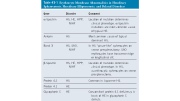
Table 43-1 Erythrocyte Membrane Abnormalities in Hereditary Spherocytosis, Hereditary Elliptocytosis, and Related Disorders
Pathobiology
Two major factors are in sic red blood cell defect a and damages abnormal dysfunction of proteins o tistep process of accelera lization of the lipid bil membrane, leading to su deformable spherocytes the spleen (see Fig. 43-2
Molecular Patholog
The molecular basis of H quantitation of membra electrophoresis, HS can isolated deficiency of sp and ankyrin, (3) defici protein 4.2, and (5) no
Isolated Spectrin D
The reported mutations of both α- and β-spect
Gene
α-Spectrin
Ankyrin
Band 3
β-Spectrin
Protein 4.2
Protein 4.1
Glycophorin C
Disorder
HS, HE, HPP, NIHF
HS
HS, SAO, NIHF
HS, HE, HPP, NIHF
HS
HE
HE
Comment
Location of mutation determines clinical phenotype. α-Spectrin mutations are most common cause of typical HE.
Most common cause of typical dominant HS.
In HS “pincer-like” spherocytes on smear presplenectomy. SAO erythrocytes have transverse ridge or longitudinal slit.
Location of mutation determines clinical phenotype. In HS, acanthrocytic spherocytes on smear presplenectomy.
Common in Japanese HS.
Concomitant protein 4.1 deficiency is basis of HE in glycophorin C defects.
