
Page 114 - Aloe Vera Information - Scientific Papers about Aloe Vera
P. 114
from the respective vendors). Electrophoresis was performed using a 12.5% separating gel and a 4%
stacking gel run in a Bio-Rad Protean II Xi vertical electrophoresis cell system at 3OmA constant
6
current . The gel was stained by silver stain kit method, and the kit was purchased from Bio-Rad. 7
Cells were grown in Dulbecco’s Minimal Medium (DMEM) supplemented with 10% heat-inactivated
horse serum, 5% fetal calf serum, 50 units penicillin, 0.05% mg/ml streptomycin 1mM L-glutamine and
o
1mM sodium pyruvate. The rat adrenal cultured cells were prepared following incubation at 37 C in 5%
CO . Cell concentration and viability were determined by hemacytometer counts and dye exclusion with
2
0.04% trypan blue.
4
Cells at 5x10 cell/well were plated into 96-well flat-bottom plates and maintained 24 hours at standard
conditions in adherence studies. The media was removed before the addition of the Aloe-DMEM mixture.
After 72 hours of incubation, 10ul of MTT (3-[4,5-Dimeth-ylthiazol-2-yl]-2,5-diphenyl-tetrazolium
bromide) solution (5mg/ml) was added to each well. The formazan crystals, formed only in viable cells
o
after four hours at 37 C, were dissolved by addition of 100ul of acid-isopropanol solution. The plates
were read by a Micro ELISA reader (MR 580 Dynatec) at 570nm (630nm reference wavelength,
calibration setting of 1.99).
8
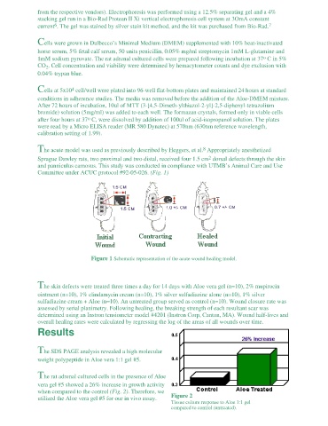
The acute model was used as previously described by Heggers, et al. Appropriately anesthetized
2
Sprague Dawley rats, two proximal and two distal, received four 1.5 cm dorsal defects through the skin
and panniculus carnosus. This study was conducted in compliance with UTMB’s Animal Care and Use
Committee under ACUC protocol #92-05-026. (Fig. 1)
Figure 1 Schematic representation of the acute wound healing model.
The skin defects were treated three times a day for 14 days with Aloe vera gel (n=10), 2% mupirocin
ointment (n=10), 1% clindamycin cream (n=10), 1% silver sulfadiazine alone (n=10), 1% silver
sulfadiazine cream + Aloe (n=10). An untreated group served as control (n=10). Wound closure rate was
assessed by serial planimetry. Following healing, the breaking strength of each resultant scar was
determined using an Instron tensiometer model #4201 (Instron Corp, Canton, MA). Wound half-lives and
overall healing rates were calculated by regressing the log of the areas of all wounds over time.
Results
The SDS PAGE analysis revealed a high molecular
weight polypeptide in Aloe vera 1:1 gel #5.
The rat adrenal cultured cells in the presence of Aloe
vera gel #5 showed a 26% increase in growth activity
when compared to the control (Fig. 2). Therefore, we
Figure 2
utilized the Aloe vera gel #5 for our in vivo assay.
Tissue culture response to Aloe 1:1 gel
compared to control (untreated).