
Page 100 - Alaska A & P Primer
P. 100
17.4 The Thyroid Gland
17.5 The Parathyroid Gland
17.4 OBJECTIVES
1. Describe the location and anatomy of the thyroid gland
17.5 OBJECTIVES
1. Describe the location and structure of the parathyroid glands
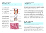
A butterfly-shaped organ, the thyroid gland is located anterior to the trachea, just inferior to the larynx. The medial region,
called the isthmus, is flanked by
wing-shaped left and right lobes.
Each of the thyroid lobes are em- bedded with parathyroid glands, primarily on their posterior sur- faces. The tissue of the thyroid gland is composed mostly of thy- roid follicles. The follicles are made up of a central cavity filled with a sticky fluid called colloid.
Surrounded by a wall of epithe- lial follicle cells, the colloid is the center of thyroid hormone pro- duction, and that production is dependent on the hormones’ es- sential and unique component: iodine.
The thyroid gland is located in the neck where it wraps around the trachea. (a) Anterior view of the thyroid gland. (b) Posterior view of the thyroid gland. (c) The glandular tissue is composed pri- marily of thyroid follicles. The larger parafollicular cells often appear within the matrix of folli- cle cells. LM X 1332. (Micrograph provided by the Regents of Uni- versity of Michigan Medical School ˝ 2012)
The parathyroid glands are tiny, round structures usually found embedded in the posterior surface of the thyroid gland. The primary functional cells of the parathyroid glands are the chief cells. These epithelial cells produce and secrete the parathyroid hormone (PTH), the major hormone involved in the regulation of blood calcium levels.
Abnormally high activity of the parathyroid gland can cause hyperparathyroidism, a disor- der caused by an overproduction of PTH that results in excessive calcium reabsorption from bone. Hyperparathyroidism can significantly decrease bone density, leading to spon- taneous fractures or deformities. As blood calcium levels rise, cell membrane permeability to sodium is decreased, and the responsiveness of the nervous system is reduced. At the same time, calcium deposits may collect in the body’s tissues and organs, impairing their functioning. In contrast, abnormally low blood calcium levels may be caused by parathy- roid hormone deficiency, called hypoparathyroidism, which may develop following injury or surgery involving the thyroid gland. Low blood calcium increases membrane permeabil- ity to sodium, resulting in muscle twitching, cramping, spasms, or convulsions. Severe defi- cits can paralyze muscles, including those involved in breathing, and can be fatal. When blood calcium levels are high, calcitonin is produced and secreted by the parafollicular
cells of the thyroid gland. A discussed earlier, calcitonin inhibits the activity of osteoclasts, reduces the absorption of dietary calcium in the intestine, and signals the kidneys to reab- sorb less calcium, resulting in larger amounts of calcium excreted in the urine.
17.6 The Adrenal Gland
The adrenal glands, located superior to each kidney, consist of two regions: the adrenal cortex and adrenal medulla. The adrenal cortex—the outer layer of the gland—produces mineralocorticoids, glucocorticoids, and androgens. The adrenal medulla at the core of the gland produces epinephrine and norepinephrine.The adrenal glands mediate a short-term stress response and a long-term stress response. A perceived threat results in the bsecre- tion of epinephrine and norepinephrine from the adrenal medulla, which mediate the fight-or-flight response. The long-term stress response is mediated by the secretion of CRH from the hypothalamus, which triggers ACTH, which in turn stimulates the secretion
17.6 OBJECTIVES
1. Describe the location and structure of the adrenal glands
This content is available for free at https://cnx.org/content/col11496/1.7
State of Alaska EMS Education Primer - 2016
99
