
Page 99 - Alaska A & P Primer
P. 99
When fetal development is complete, the peptide-derived hormone oxytocin (tocia- = “childbirth”) stimulates uterine contractions and dilation of the cervix. Throughout most of pregnancy, oxytocin hormone receptors are not expressed at high levels in the uterus. Toward the end of pregnancy, the synthesis of oxytocin receptors in the uterus increases, and the smooth muscle cells of the uterus become more sensitive to its effects. Oxytocin is also thought to be involved in feelings of love and closeness, as well as in the sexual re- sponse.
In response to high blood osmolarity, which can occur during dehydration or following a very salty meal, the osmoreceptors signal the posterior pituitary to release antidiuretic hor- mone (ADH). The target cells of ADH are located in the tubularmore water reabsorbed from the filtrate, the greater the amount of water that is returned to the blood and the less that is excreted in the urine. A greater concentration of water results in a reduced concen- tration of solutes. ADH is also known as vasopressin because, in very high concentrations, it causes constriction of blood vessels, which increases blood pressure by increasing periph- eral resistance.
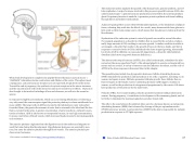
The anterior pituitary originates from the digestive tract in the embryo and migrates to- ward the brain during fetal development. Hypothalamic hormones are secreted by neu- rons, but enter the anterior pituitary through blood vessels. The anterior pituitary pro- duces seven hormones.
The endocrine system regulates the growth of the human body, protein synthesis, and cel- lular replication. A major hormone involved in this process is growth hormone (GH), also called somatotropin—a protein hormone produced and secreted by the anterior pituitary gland. Its primary function is anabolic; it promotes protein synthesis and tissue building through direct and indirect mechanisms.
A glucose-sparing effect occurs when GH stimulates lipolysis, or the breakdown of adipose tissue, releasing fatty acids into the blood. As a result, many tissues switch from glucose to fatty acids as their main energy source, which means that less glucose is taken up from the bloodstream.
Dysfunction of the endocrine system’s control of growth can result in several disorders. For example, gigantism is a disorder in children that is caused by the secretion of abnor- mally large amounts of GH, resulting in excessive growth. A similar condition in adults is acromegaly, a disorder that results in the growth of bones in the face, hands, and feet in response to excessive levels of GH in individuals who have stopped growing. Abnormally low levels of GH in children can cause growth impairment—a disorder called pituitary dwarfism (also known as growth hormone deficiency).
The adrenocorticotropic hormone (ACTH), also called corticotropin, stimulates the adre- nal cortex (the more superficial “bark” of the adrenal glands) to secrete corticosteroid hor- mones such as cortisol. A variety of stressors can also influence its release, and the role of ACTH in the stress response is discussed later in this chapter.
The gonadotropins include two glycoprotein hormones: follicle-stimulating hormone (FSH) stimulates the production and maturation of sex cells, or gametes, including ova in women and sperm in men. FSH also promotes follicular growth; these follicles then re- lease estrogens in the female ovaries. Luteinizing hormone (LH) triggers ovulation in women, as well as the production of estrogens and progesterone by the ovaries. LH stimu- lates production of testosterone by the male testes.
Prolactin (PRL): As its name implies, prolactin promotes lactation (milk production) in women. During pregnancy, it contributes to development of the mammary glands, and af- ter birth, it stimulates the mammary glands to produce breast milk.
The cells in the zone between the pituitary lobes secrete a hormone known as melanocyte- stimulating hormone (MSH) that is formed by cleavage of the pro-opiomelanocortin (POMC) precursor protein. Local production of MSH in the skin is responsible for melanin production in response to UV light exposure.
This content is available for free at https://cnx.org/content/col11496/1.7
State of Alaska EMS Education Primer - 2016
98
