
Page 111 - Alaska A & P Primer
P. 111
The middle and thickest layer is the myocar- dium, made largely of cardiac muscle cells. It is built upon a framework of collagenous fibers, plus the blood vessels that supply the myocar- dium and the nerve fibers that help regulate the heart. It is the contraction of the myocar- dium that pumps blood through the heart and into the major arteries.
Although the ventricles on the right and left sides pump the same amount of blood per con- traction, the muscle of the left ventricle is much thicker and better developed than that of the right ventricle. In order to overcome the high resistance required to pump blood into
the long systemic circuit, the left ventricle must generate a great amount of pressure. The right ventricle does not need to generate as much pressure, since the pulmonary circuit is shorter and provides less resistance.
The innermost layer of the heart wall, the endocardium, is joined to the myocardium with a thin layer of connective tissue. The endocardium lines the chambers where the blood cir- culates and covers the heart valves. It is made of simple squamous epithelium called endo- thelium, which is continuous with the endothelial lining of the blood vessels
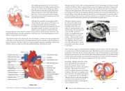
The right atrium serves as the receiving chamber for blood returning to the heart from the systemic circulation. The two major systemic veins, the superior and inferior venae cavae, and the large coronary vein called the coronary sinus that drains the heart myocardium empty into the right atrium. The atria receive venous blood on a nearly continuous basis, preventing venous flow from stopping while the ventricles are contracting. While most ven- tricular filling occurs while the atria are relaxed, they do demonstrate a contractile phase and actively pump blood into the ventricles just prior to ventricular contraction. The open- ing between the atrium and ventricle is guarded by the tricuspid valve.
The right ventricle receives blood from the right atrium through the tricuspid valve. Each flap of the valve is at- tached to strong strands of con- nective tissue, the chordae tend- ineae, literally “tendinous cords,” or sometimes more po- etically referred to as “heart strings.” There are three papil- lary muscles in the right ventri- cle, called the anterior, poste- rior, and septal muscles, which correspond to the three sections of the valves.
After exchange of gases in the pulmonary capillaries, blood returns to the left atrium high in oxygen via one of the four pulmonary veins. Blood flows nearly continuously from the pulmonary veins back into the atrium, which acts as the receiving chamber, and from here through an opening into the left ventricle. Most blood flows passively into the heart while both the atria and ventricles are relaxed, but toward the end of the ventricular relaxation period, the left atrium will contract, pumping blood into the ventricle.
Recall that, although both sides of the heart will pump the same amount of blood, the muscular layer is much thicker in the left ventricle compared to the right. The mitral valve is con- nected to papillary muscles via chordae tendineae. There are two papillary mus- cles on the left—the anterior and poste- rior—as opposed to three on the right. The left ventricle is the major pumping chamber for the systemic circuit; it ejects blood into the aorta through the aortic semilunar valve.
This content is available for free at https://cnx.org/content/col11496/1.7
State of Alaska EMS Education Primer - 2016
110
