
Page 112 - Alaska A & P Primer
P. 112
Coronary veins drain the heart and generally parallel the large surface arteries. The great cardiac vein can be seen initially on the surface of the heart following the interventricular sulcus, but it eventually flows along the coronary sulcus into the coronary sinus on the pos- terior surface. The great cardiac vein initially parallels the anterior interventricular artery and drains the areas supplied by this vessel. It receives several major branches, including the posterior cardiac vein, the middle cardiac vein, and the small cardiac vein.
The posterior cardiac vein parallels and drains the areas supplied by the marginal artery branch of the circumflex artery. The middle cardiac vein parallels and drains the areas sup- plied by the posterior interventricular artery. The small cardiac vein parallels the right coronary artery and drains the blood from the posterior surfaces of the right atrium and ventricle. The coronary sinus is a large, thin-walled vein on the posterior surface of the heart lying within the atrioventricular sulcus and emptying directly into the right atrium. The anterior cardiac veins parallel the small cardiac arteries and drain the anterior surface of the right ventricle. Unlike these other cardiac veins, it bypasses the coronary sinus and drains directly into the right atrium.
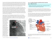
Coronary artery disease is the leading cause of death worldwide. It occurs when the buildup of plaque—a fatty material including cholesterol, connective tissue, white blood cells, and some smooth muscle cells—within the walls of the arteries obstructs the flow of blood and decreases the flexibility or compliance of the vessels. This condition is called atherosclerosis, a hardening of the arteries that involves the accumulation of plaque. As the coronary blood ves-
sels become occluded,
the flow of blood to the
tissues will be re-
stricted, a condition
called ischemia that
causes the cells to re-
ceive insufficient
amounts of oxygen,
called hypoxia. This
picture shows the
blockage of coronary
arteries highlighted by
the injection of dye.
Some individuals with
coronary artery dis-
ease report pain radiat-
ing from the chest
called angina pectoris,
but others remain as-
ymptomatic. If un-
treated, coronary ar-
tery disease can lead to
MI or a heart attack.
19.2 Cardiac Muscle and Electrical Activity
Recall that cardiac muscle shares a few characteristics with both skeletal muscle and smooth muscle, but it has some unique properties of its own. Not the least of these excep- tional properties is its ability to initiate an electrical potential at a fixed rate that spreads rapidly from cell to cell to trigger the contractile mechanism. This property is known as autorhythmicity.
There are two major types of cardiac muscle cells: myocardial contractile cells and myocar- dial conducting cells. Themyocardial contractile cells constitute the bulk (99 percent) of the cells in the atria and ventricles. Contractile cells conductimpulses and are responsible for contractions that pump blood through the body. The myocardial conducting cells (1 percent of the cells) form the conduction system of the heart.
The components of the cardiac conduction system include the sinoatrial node, the atrioven- tricular node, the atrioventricular bundle, the atrioventricular bundle branches, and the Purkinje cells.
19.2 OBJECTIVES
1. Identify and describe the components of the conducting system that distributes electrical impulses through the heart
This content is available for free at https://cnx.org/content/col11496/1.7
State of Alaska EMS Education Primer - 2016
111
