
Page 135 - Alaska A & P Primer
P. 135
constant beating motion, sweeping materials towards the throat to be swallowed. Interest- ingly, cold air slows the movement of the cilia, resulting in accumulation of mucus that may in turn lead to a runny nose during cold weather. This moist epithelium functions to warm and humidify incoming air. Capillaries located just beneath the nasal epithelium warm the air by convection. Serous and mucus-producing cells also secrete the lysozyme enzyme and proteins called defensins, which have antibacterial properties. Immune cells that patrol the connective tissue deep to the respiratory epithelium provide additional pro- tection.
The pharynx is divided into three major regions: the na- sopharynx, the oropharynx, and the laryngopharynx.
A pharyngeal tonsil, also called an adenoid, is an ag- gregate of lymphoid reticu- lar tissue similar to a lymph node that lies at the superior portion of the nasopharynx. The function of the pharyn- geal tonsil is not well under- stood, but it contains a rich supply of lymphocytes and is covered with ciliated epithe-
lium that traps and destroys invading pathogens that enter during inhalation. The pharyn- geal tonsils are large in children, but interestingly, tend to regress with age and may even disappear. The oropharynx is a passageway for both air and food. The oropharynx is bor- dered superiorly by the nasopharynx and anteriorly by the oral cavity. The fauces is the opening at the connection between the oral cavity and the oropharynx. As the nasopharynx becomes the oropharynx, the epithelium changes from pseudostratified ciliated columnar epithelium to stratified squamous epithelium. The oropharynx contains two distinct sets of tonsils, the palatine and lingual tonsils.
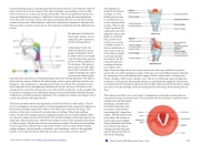
The larynx extends from the laryngopharynx and the hyoid bone to the trachea. The lar- ynx is a cartilaginous structure inferior to the laryngopharynx that connects the pharynx to the trachea and helps regulate the volume of air that enters and leaves the lungs. The structure of the larynx is formed by several pieces of cartilage. Three large cartilage pieces—the thyroid cartilage (anterior), epiglottis (superior), and cricoid cartilage (inferi- or)—form the major structure of the larynx. The thyroid cartilage is the largest piece of car- tilage that makes up the larynx. The thyroid cartilage consists of the laryngeal prominence, or “Adam’s apple,” which tends to be more prominent in males. The thick cricoid cartilage forms a ring, with a wide posterior region and a thinner anterior region. Three smaller, paired cartilages—the arytenoids, corniculates, and cuneiforms—attach to the epiglottis and the vocal cords and muscle that help move the vocal cords to produce speech.
The epiglottis, at-
tached to the thyroid
cartilage, is a very
flexible piece of elas-
tic cartilage that cov-
ers the opening of the
trachea. When in the
“closed” position, the
unattached end of the
epiglottis rests on the
glottis. A vestibular
fold, or false vocal
cord, is one of a pair
of folded sections of
mucous membrane. A
true vocal cord is one
of the white, membra-
nous folds attached
by muscle to the thy-
roid and arytenoid
cartilages of the lar-
ynx on their outer
edges. The inner edges of the true vocal cords are free, allowing oscillation to produce sound. The size of the membranous folds of the true vocal cords differs between individu- als, producing voices with different pitch ranges. Folds in males tend to be larger than those in females, which create a deeper voice. The act of swallowing causes the pharynx and larynx to lift upward, allowing the pharynx to expand and the epiglottis of the larynx to swing downward, closing the opening to the trachea. These movements produce a larger area for food to pass through, while preventing food and beverages from entering the tra- chea.
The trachea is formed by 16 to 20 stacked, C-shaped pieces of hyaline cartilage that are connected by dense connective tissue. The trachealis muscle and elastic connective tissue together form the fibroelastic
membrane, a flexible mem-
brane that closes the poste- rior surface of the trachea, connecting the C-shaped car- tilages. The fibroelastic mem- brane allows the trachea to stretch and expand slightly during inhalation and exhala- tion, whereas the rings of cartilage provide structural support and prevent the tra- chea from collapsing.
This content is available for free at https://cnx.org/content/col11496/1.7
State of Alaska EMS Education Primer - 2016
134
