
Page 137 - Alaska A & P Primer
P. 137
22.2 The Lungs
A major organ of the respiratory system, each lung houses structures of both the conduct- ing and respiratory zones. The main function of the lungs is to perform the exchange of oxygen and carbon dioxide with air from the atmosphere. To this end, the lungs exchange respiratory gases across a very large epithelial surface area—about 70 square meters—that is highly permeable to gases.
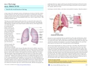
The lungs are pyramid-shaped, paired organs that are connected to the trachea by the right and left bronchi; on the inferior surface, the lungs are bordered by the diaphragm. The diaphragm is the flat, dome-shaped muscle located at the base of the lungs and tho- racic cavity. The lungs
are enclosed by the pleurae, which are at- tached to the mediasti- num. The right lung is shorter and wider than the left lung, and the left lung occupies a smaller volume than the right. The cardiac notch is an indentation on the surface of the left lung, and it allows space for the heart.
The blood supply of the
lungs plays an impor-
tant role in gas exchange
and serves as a transport system for gases throughout the body. In addition, innervation by the both the parasympathetic and sympathetic nervous systems provides an important level of control through dilation and constriction of the airway. The major function of the lungs is to perform gas exchange, which requires blood from the pulmonary circulation. This blood supply contains deoxygenated blood and travels to the lungs where erythro- cytes, also known as red blood cells, pick up oxygen to be transported to tissues through- out the body. Dilation and constriction of the airway are achieved through nervous con- trol by the parasympathetic and sympathetic nervous systems. The parasympathetic sys- tem causes bronchoconstriction, whereas the sympathetic nervous system stimulates bron- chodilation. Reflexes such as coughing, and the ability of the lungs to regulate oxygen and carbon dioxide levels, also result from this autonomic nervous system control. Sensory nerve fibers arise from the vagus nerve, and from the second to fifth thoracic ganglia. The
pulmonary plexus is a region on the lung root formed by the entrance of the nerves at the hilum. The nerves then follow the bronchi in the lungs and branch to innervate muscle fi- bers, glands, and blood vessels.
Each lung is enclosed within a cavity that is surrounded by the pleura. The pleura (plural =
pleurae) is a serous membrane that surrounds the lung. The right and left pleurae, which enclose the right and left lungs, respectively, are separated by the mediastinum. The pleu- rae consist of two layers. The visceral pleura is the layer that is superficial to the lungs, and extends into and lines the lung fissures. In contrast, the parietal pleura is the outer layer that connects to the thoracic wall, the mediastinum, and the diaphragm.
The visceral and parietal pleurae connect to each other at the hilum. The pleural cavity is the space between the visceral and parietal layers. The pleurae perform two major func- tions: They produce pleural fluid and create cavities that separate the major organs. Pleu- ral fluid is secreted by mesothelial cells from both pleural layers and acts to lubricate their surfaces. This lubrication reduces friction between the two layers to prevent trauma during breathing, and creates surface tension that helps maintain the position of the lungs against the thoracic wall. This adhesive characteristic of the pleural fluid causes the lungs to en- large when the thoracic wall expands during ventilation, allowing the lungs to fill with air. The pleurae also create a division between major organs that prevents interference due to the movement of the organs, while preventing the spread of infection.
22.2 OBJECTIVES
1. Describe the overall function of the lung
Listen to Lungs sounds:
http://www.littmann.ca/wps/portal/3M/en_CA/3M-Littmann-CA/stethoscope/littman
n-learning-institute/heart-lung-sounds/lung-sounds/
This content is available for free at https://cnx.org/content/col11496/1.7
State of Alaska EMS Education Primer - 2016
136
