
Page 43 - Alaska A & P Primer
P. 43
7.4 The Thoracic Cage
7.5 Embryonic Development of the Axial Skeleton
7.4 OBJECTIVES
1. Describe the components that make up the thoracic cage
7.5 OBJECTIVES
1. Describe the two types of embryonic bone development within the skull
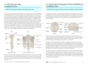
The thoracic cage protects the heart and lungs. It is composed of 12 pairs of ribs with their costal cartilages and the sternum. The ribs are anchored posteriorly to the 12 thoracic ver- tebrae. The sternum consists of the manubrium, body, and xiphoid process. The manu- brium and body are joined at the sternal angle, which is also the site for attachment of the second ribs. Ribs are flattened, curved bones and are numbered 1–12. Posteriorly, the head of the rib articulates with the costal facets located on the bodies of thoracic vertebrae and the rib tubercle articulates with the facet located on the vertebral transverse process. The angle of the ribs forms the most posterior portion of the thoracic cage. The costal groove in the inferior margin of each rib carries blood vessels and a nerve. Anteriorly, each rib ends in a costal cartilage. True ribs (1–7) attach directly to the sternum via their costal cartilage. The false ribs (8–12) either attach to the sternum indirectly or not at all. Ribs 8– 10 have their costal cartilages attached to the cartilage of the next higher rib. The floating ribs (11–12) are short and do not attach to the sternum or to another rib.
Thoracic Cage The thoracic cage is formed by the (a) sternum and (b) 12 pairs of ribs with their costal cartilages. The ribs are anchored posteriorly to the 12 thoracic vertebrae. The sternum consists of the manubrium, body, and xiphoid process. The ribs are classified as true ribs (1–7) and false ribs (8–12). The last two pairs of falseribs are also known as float- ing ribs (11–12).
The axial skeleton begins to form during early embryonic development. However, growth, remodeling, and ossification (bone formation) continue for several decades after birth be- fore the adult skeleton is fully formed. Knowledge of the developmental processes that give rise to the skeleton is important for understanding the abnormalities that may arise in skeletal structures.
The bones of the newborn skull are not fully ossified and are separated by large areas called fontanelles, which are filled with fibrous connective tissue. The fontanelles allow for continued growth of the skull after birth. At the time of birth, the facial bones are small and underdeveloped, and the mastoid process has not yet formed.
Development of the vertebrae begins with the accumulation of mesenchyme cells from each sclerotome around the notochord. These cells differentiate into a hyaline cartilage model for each vertebra, which then grow and eventually ossify into bone through the proc- ess of endochondral ossification. As the developing vertebrae grow, the notochord largely disappears. However, small areas of notochord tissue persist between the adjacent verte- brae and this contributes to the formation of each intervertebral disc.
The ribs and sternum also develop from mesenchyme. The ribs initially develop as part of the cartilage model for each vertebra, but in the thorax region, the rib portion separates from the vertebra by the eighth week. The cartilage model of the rib then ossifies, except for the anterior portion, which remains as the costal cartilage. The sternum initially forms as paired hyaline cartilage models on either side of the anterior midline, beginning during the fifth week of development.
This content is available for free at https://cnx.org/content/col11496/1.7
State of Alaska EMS Education Primer - 2016
42
