
Page 46 - Alaska A & P Primer
P. 46
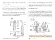
The ulna is the medial bone of the forearm. It runs parallel to the radius, which is the lat- eral bone of the forearm. The proximal end of the ulna resembles a crescent wrench with its large, C-shaped trochlear notch. This region articulates with the trochlea of the hu- merus as part of the elbow joint.
More distal is the shaft of the ulna. The lateral side of the shaft forms a ridge called the in- terosseous border of the ulna. This is the line of attachment for the interosseous mem- brane of the forearm, a sheet of dense connective tissue that unites the ulna and radius bones. The small, rounded area that forms the distal end is the head of the ulna. Projecting from the posterior side of the ulnar head is the styloid process of the ulna, a short bony projection. This serves as an attachment point for a connective tissue structure that unites the distal ends of the ulna and radius.
In the anatomical position, with the elbow fully extended and the palms facing forward, the arm and forearm do not form a straight line. Instead, the forearm deviates laterally by 5–15 degrees from the line of the arm. This deviation is called the carrying angle. It allows the forearm and hand to swing freely or to carry an object without hitting the hip. The car- rying angle is larger in females to accommodate their wider pelvis.
The radius runs parallel to the ulna, on the lateral (thumb) side of the forearm. The head of the radius is a disc-shaped structure that forms the proximal end. The small depression on the surface of the head articulates with the capitulum of the humerus as part of the el- bow joint, whereas the smooth, outer margin of the head articulates with the radial notch of the ulna at the proximal radioulnar joint. The neck of the radius is the narrowed region immediately below the expanded head. Inferior to this point on the medial side is the ra- dial tuberosity, an oval-shaped, bony protuberance that [is] a muscle attachment point.
Bones of the Wrist and Hand
The eight carpal bones form the base of the hand. These are arranged into proximal and distal rows of four bones each. The metacarpal bones form the palm of the hand. The thumb and fingers consist of the phalanx bones. Within the carpal bones, the four proxi- mal bones are united to each other by ligaments to form a unit. Only three of these bones, the scaphoid, lunate, and triquetrum, contribute to the radiocarpal joint.
The four distal carpal bones are also held together as a group by ligaments. The proximal and distal rows of carpal bones articulate with each other to form the midcarpal joint. To- gether, the radiocarpal and midcarpal joints are responsible for all movements of the hand at the wrist. The distal carpal bones also articulate with the metacarpal bones of the hand.
Visit this site (http://openstaxcollege.org/l/handbone) to explore the bones and joints of the hand. What are the three arches of the hand, and what is the importance of these during the gripping of an object?
This content is available for free at https://cnx.org/content/col11496/1.7
State of Alaska EMS Education Primer - 2016
45
