
Page 49 - Alaska A & P Primer
P. 49
8.4 Bones of the Lower Limb
8.4 OBJECTIVES
1. Describe the bones and bony landmarks that articulate at each joint of the lower limb
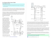
The lower limb is divided into three regions. These are the thigh, located between the hip and knee joints; the leg, located between the knee and ankle joints; and distal to the ankle, the foot. There are 30 bones in each lower limb. These are the femur, patella, tibia, fibula, seven tarsal bones, five metatarsal bones, and 14 phalanges.
The femur is the single bone of the thigh region. It articulates superiorly with the hip bone at the hip joint, and inferiorly with the tibia at the knee joint. The patella only articulates with the distal end of the femur. The narrowed region below the head is the neck of the femur. This is a common area for fractures of the femur. The greater trochanter is the large, upward, bony projection
located above the base of the neck.
Anteriorly, the smooth sur- faces of the condyles join to- gether to form a wide groove called the patellar surface, which provides for articula- tion with the patella bone. The combination of the medial and lateral condyles with the patel- lar surface gives the distal end of the femur a horseshoe (U) shape. The leg contains the large tibia on the medial side and the slender fibula on the lateral side. The tibia bears
the weight of the body, whereas the fibula does not bear weight. The interosseous border of each bone is the at- tachment site for the interosse- ous membrane of the leg, the connective tissue sheet that unites the tibia and fibula.
The tibial tuberosity is an ele- vated area on the anterior side
of the tibia, near its proximal end. It is the final site of attachment for the muscle tendon associated with the patella. More inferiorly, the shaft of the tibia becomes triangular in shape. Both the anterior border and the medial side of the triangular shaft are located im- mediately under the skin and can be easily palpated along the entire length of the tibia.
These details provide easy access for intraosseous cannulation. A small ridge running down the lateral side of the tibial shaft is the interosseous border of the tibia.
The fibula is the slender bone located on the lateral side of the leg. The fibula does not bear weight. It serves primarily for muscle attachments and thus is largely surrounded by mus- cles. Only the proximal and distal ends of the fibula can be palpated. The head of the fib- ula is the small, knob-like, proximal end of the fibula. It articulates with the inferior aspect of the lateral tibial condyle, forming the proximal tibiofibular joint.
The thin shaft of the fibula has the interosseous border of the fibula, a narrow ridge run- ning down its medial side for the attachment of the interosseous membrane that spans the fibula and tibia.
Watch video of real emergency intraosseous cannulation of the tibial tuberosity:
https://www.youtube.com/watch?v=UXVDx26N9Zk
This content is available for free at https://cnx.org/content/col11496/1.7
State of Alaska EMS Education Primer - 2016
48
