
Page 50 - Alaska A & P Primer
P. 50
The posterior half of the foot is formed by seven tarsal bones. The most superior bone is the talus. This has a relatively square-shaped, upper surface that articulates with the tibia and fibula to form the ankle joint. Three areas of articulation form the ankle joint: The su- peromedial surface of the talus bone articulates with the medial malleolus of the tibia, the top of the talus articulates with the distal end of the tibia, and the lateral side of the talus articulates with the lateral malleolus of the fibula.
Inferiorly, the talus articulates with the calcaneus (heel bone), the largest bone of the foot, which forms the heel. Body weight is transferred from the tibia to the talus to the cal- caneus, which rests on the ground. The medial calcaneus has a prominent bony extension called the sustentaculum tali (“support for the talus”) that supports the medial side of the talus bone.
The anterior half of the foot is formed by the five metatarsal bones, which are located be- tween the tarsal bones of the posterior foot and the phalanges of the toes. These elongated bones are numbered 1–5, starting with the medial side of the foot. Each metatarsal bone articulates with the proximal phalanx of a toe to form a metatarsophalangeal joint. The heads of the metatarsal bones also rest on the ground and form the ball (anterior end) of the foot.
The toes contain a total of 14 phalanx bones (phalanges), arranged in a similar manner as the phalanges of the fingers. The toes are numbered 1–5, starting with the big toe ( hallux). The big toe has two phalanx bones, the proximal and distal phalanges. The remaining toes
all have proximal, middle, and distal phalanges. A joint between adjacent phalanx bones is called an interphalangeal joint.
When the foot comes into contact with the ground during walking, running, or jumping activities, the impact of the body weight puts a tremendous amount of pressure and force on the foot. During running, the force applied to each foot as it contacts the ground can be up to 2.5 times your body weight. The bones, joints, ligaments, and muscles of the foot ab- sorb this force, thus greatly reducing the amount of shock that is passed superiorly into the lower limb and body.
The arches ofthe foot play an important role in this shock-absorbing ability. When weight is applied to the foot, these arches will flatten somewhat, thus absorbing energy. When the weight is removed, the arch rebounds, giving “spring” to the step. The arches also serve to distribute body weight side to side and to either end of the foot.
Stretching of the ligaments that support the longitudinal arches can lead to pain. This can occur in overweight individuals, with people who have jobs that involve standing for long periods of time (such as a waitress), or walking or running long distances. If stretching of the ligaments is prolonged, excessive, or repeated, it can result in a gradual lengthening of the supporting ligaments, with subsequent depression or collapse of the longitudinal arches, particularly on the medial side of the foot. This condition is called pes planus (“flat foot” or “fallen arches”).
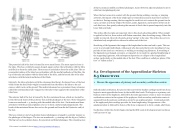
8.5 Development of the Appendicular Skeleton
Endochondral ossification, the process that converts the hyaline cartilage model into bone, begins in most appendicular bones by the twelfth fetal week. This begins as a primary ossi- fication center in the diaphysis, followed by the later appearance of one or more secondary ossifications centers in the regions of the epiphyses. Each secondary ossification center is separated from the primary ossification center by an epiphyseal plate. Continued growth of the epiphyseal plate cartilage provides for bone lengthening. Disappearance of the epiphyseal plate is followed by fusion of the bony components to form a single, adult bone.
Ossification within the clavicle begins during the fifth week of development and continues until 25 years of age.
8.5 OBJECTIVES
1. Discuss the appearance of primary and secondary ossification centers
This content is available for free at https://cnx.org/content/col11496/1.7
State of Alaska EMS Education Primer - 2016
49
