
Page 58 - Alaska A & P Primer
P. 58
The medial elbow is supported by the ulnar collateral ligament and the radial collateral ligament supports the lateral side. These ligaments pre- vent side-to-side movements and resist hyperextension of the elbow.
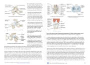
The hip joint is a ball-and-socket joint whose motions are more re- stricted than at the shoulder to pro- vide greater stability during weight bearing. The hip joint is the articula- tion between the head of the femur and the acetabulum of the hip bone.
The acetabulum is deepened by the acetabular labrum. The iliofemoral, pubofemoral, and ischiofemoral liga- ments strongly support the hip joint in the upright, standing position. The ligament of the head of the fe- mur provides little support but car- ries an important artery that sup- plies the femur.
The knee includes three articula- tions. The femoropatellar joint is between the patella and distal fe- mur. The patella, asesamoid bone incorporated into the tendon of the quadriceps femoris muscle of the anterior thigh, serves to protect this tendon from rubbing against the dis-
tal femur during knee movements. During these movements, the condyles of the femur both roll and glide over the surface of
the tibia. As the knee comes into full extension, a slight medial rotation of the femur serves to “lock” the knee into its most stable, weight-bearing position.
Injuries to the knee are common. Since this joint is primarily supported by muscles and ligaments, injuries to any of these structures will result in pain or knee instability. Injury to the posterior cruciate ligament occurs when the knee is flexed and the tibia is driven posteriorly, such as falling and landing on the tibial tuberosity or hitting the tibia on the dashboard when not wearing a seatbelt during an automobile accident. More commonly, injuries occur when forces are applied to the extended knee, particularly when the foot is planted and unable to move. Anterior cruciate ligament injuries can result with a forceful
blow to the anterior knee, producing hyperextension, or when a runner makes a quick change of direction that produces both twisting and hyperextension of the knee.
A worse combination of injuries can occur with a hit to the lateral side of the extended knee. A moderate blow to the lateral knee will cause the medial side of the joint to open, resulting in stretching or damage to the tibial collateral ligament. Because the medial me- niscus is attached to the tibial collateral ligament, a stronger blow can tear the ligament and also damage the medial meniscus. This is one reason that the medial meniscus is 20 times more likely to be injured than the lateral meniscus. A powerful blow to the lateral knee produces a “terrible triad” injury, in which there is a sequential injury to the tibial collateral ligament, medial meniscus, and anterior cruciate ligament.
Arthroscopic surgery has greatly improved the surgical treatment of knee injuries and re- duced subsequent recovery times. This procedure involves a small incision and the inser- tion into the joint of an arthroscope, a pencil-thin instrument that allows for visualization of the joint interior. Small surgical instruments are also inserted via additional incisions. These tools allow a surgeon to remove or repair a torn meniscus or to reconstruct a rup- tured cruciate ligament. The current method for anterior cruciate ligament replacement involves using a portion of the patellar ligament. Holes are drilled into the cruciate liga- ment attachment points on the tibia and femur, and the patellar ligament graft, with small areas of attached bone still intact at each end, is inserted into these holes. The bone-to- bone sites at each end of the graft heal rapidly and strongly, thus allowing rapid recovery.
This content is available for free at https://cnx.org/content/col11496/1.7
State of Alaska EMS Education Primer - 2016
57
