
Page 60 - Alaska A & P Primer
P. 60
10.1 Overview of Muscle Tissues
Muscle is one of the four primary tissue types of the body, and the body contains three types of muscle tissue: skeletal muscle, cardiac muscle, and smooth muscle. All three muscle tissues have some properties in common; they all exhibit a quality called excitabil- ity as their plasma membranes can change their electrical states (from polarized to depolar- ized) and send an electrical wave called an action potential along the entire length of the membrane. While the nervous system can influence the excitability of cardiac and smooth muscle to some degree, skeletal muscle completely depends on signaling from the nervous system to work properly. On the other hand, both cardiac muscle and smooth muscle can respond to other stimuli, such as hormones and local stimuli.
The muscles all begin the actual process of contracting (shortening) when a protein called actin is pulled by a protein called myosin. This occurs in striated uscle (skeletal and car- diac) after specific binding sites on the actin have been exposed in response to the interac- tion between calcium ions (Ca++) and proteins (troponin and tropomyosin) that “shield” the actin-binding sites. Ca++ also is required for the contraction of smooth muscle, al- though its role is different: here Ca++ activates enzymes, which in turn activate myosin heads. All muscles require adenosine triphosphate (ATP) to continue the process of con- tracting, and they all relax when the Ca++ is removed and the actin-binding sites are re- shielded. A muscle can return to its original length when relaxed due to a quality of mus- cle tissue called elasticity. It can recoil back to its original length due to elastic fibers. Mus- cle tissue also has the quality of extensibility; it can stretch or extend. Contractility allows muscle tissue to pull on its attachment points and shorten with force. Differences among the three muscle types include
the microscopic organization of their contractile proteins—actin and myosin. The actin and my- osin proteins are arranged very regularly in the cytoplasm of individual muscle cells (referred to as fibers) in both skeletal mus- cle and cardiac muscle, which creates a pattern, or stripes, called striations. The striations are visible with a light micro- scope under high magnification. Skeletal muscle fibers are multi- nucleated structures that com- pose the skeletal muscle. Car-
diac muscle fibers each have one to two nuclei and are physically and electrically con- nected to each other so that the entire heart contracts as one unit (called a syncytium). Be- cause the actin and myosin are not arranged in such regular fashion in smooth muscle, the cytoplasm of a smooth muscle fiber (which has only a single nucleus) has a uniform, non- striated appearance (resulting in the name
smooth muscle). However, the less organ-
ized appearance of smooth muscle should
not be interpreted as less efficient.
10.1 OBJECTIVES
1. Describe the different types of muscle and their contractibility and extensibility
Smooth muscle in the walls of arteries is a critical component that regulates blood pressure necessary to push blood through the circulatory system; and smooth muscle in the skin, visceral organs, and internal passageways is essential for moving all ma- terials through the body.
10.2 Skeletal Muscle
Watch https://youtu.be/bwOE1MEginA
MOVIE 1.21 Three Types of Muscle 11:28 minutes Khan Academy
10.2 OBJECTIVES
1. Identify areas of the skeletal muscle fibers
Skeletal muscles contain connective tissue, blood vessels, and nerves. There are three lay- ers of connective tissue: epimysium, perimysium, and endomysium. Skeletal muscle fibers are organized into groups called fascicles. Blood vessels and nerves enter the connective tissue and branch in the cell. Muscles attach to bones directly or through tendons or apo- neuroses. Skeletal muscles maintain posture, stabilize bones and joints, control internal movement, and generate heat.
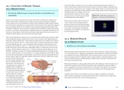
Skeletal muscle fibers are long, multinucleated cells. The membrane of the cell is the sarcol- emma; the cytoplasm of the cell is the sarcoplasm. The sarcoplasmic reticulum (SR) is a form of endoplasmic reticulum. Muscle fibers are composed of myofibrils. The striations are created by the organization of actin and myosin resulting in the banding pattern of myofibrils.
A skeletal muscle fiber is surrounded by a plasma membrane called the sarcolemma, which contains sarcoplasm, the cytoplasm of muscle cells. A muscle fiber is composed of many fibrils, which give the cell its striated appearance.
This content is available for free at https://cnx.org/content/col11496/1.7
State of Alaska EMS Education Primer - 2016
59
