
Page 45 - Tobillo y Pie 9.1
P. 45
Villena DS, Benedetti EL, Fiorentini GA
angulación del paciente en la camilla, ser sintetizadas en
forma estable por vía medial.
Por último, al estar el abordaje sobre el borde posterior
del peroné nos ofrece la ventaja de alejarnos del trayecto
del nervio sural que habitualmente se encuentra en un
punto 7cm proximal a punta del peroné, a una distancia
entre 15 a 30mm del borde posterior del mismo. De
(8)
todas maneras, debido a las variaciones anatómicas en
el curso del nervio, recomendamos la disección roma
de los tejidos circundantes.
BIBLIOGRAFÍA
1. Court-Brown CM, McBirnie J, Wilson G. Adult ankle fractures-an
increasing problem? Acta Orthop Scand. 1998;69(1):43-7.
2. Tejwani NC, Pahk B, Egol KA. Effect of posterior malleolus
fracture on outcome after unstable ankle fracture. J Trauma.
2010;69(3):666-9.
3. Harper MC, Hardin G. Posterior malleolar fractures of the ankle
associated with external rotation-abduction injuries. Results
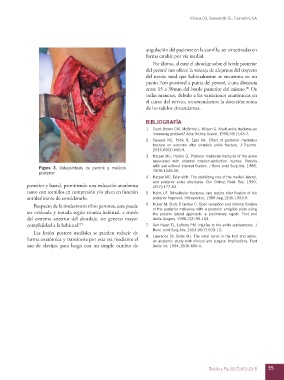
Figura 3. Osteosíntesis de peroné y maléolo with and without internal fixation. J Bone Joint Surg Am. 1988;
posterior 70(9):1348-56.
4. Harper MC. Talar shift. The stabilizing role of the medial, lateral,
and posterior ankle structures. Clin Orthop Relat Res. 1990;
posterior y lateral, permitiendo una reducción anatómica (257):177-83.
tanto con tornillos en compresión y/o placa en función 5. Heim UF. Trimalleolar fractures: late results after fixation of the
antideslizante de considerarlo. posterior fragment. Orthopedics. 1989 Aug;12(8):1053-9.
Respecto de la sindesmosis tibio peronea, esta puede 6. Huber M, Stutz P, Gerber C. Open reduction and internal fixation
ser evaluada y tratada según técnica habitual, a través of the posterior malleolus with a posterior antiglide plate using
the postero lateral approach: a preliminary report. Foot and
del extremo anterior del abordaje, sin generar mayor Ankle Surgery. 1996;2(2):95-103.
complejidad a la habitual. (7) 7. Van Heest TJ, Lafferty PM. Injuries to the ankle syndesmosis. J
Las lesión postero mediales se pueden reducir de Bone Joint Surg Am. 2014;96(7):603-13.
forma anatómica y transitoria por esta vía mediante el 8. Lawrence SJ, Botte MJ. The sural nerve in the foot and ankle:
an anatomic study with clinical and surgical implications. Foot
uso de clavijas, para luego con un simple cambio de Ankle Int. 1994;15(9):490-4.
Tobillo y Pie 2017;9(1):33-5 35