
Page 17 - Brain PET
P. 17
Marcus et al. Page 17
NIH-PA Author Manuscript
NIH-PA Author Manuscript
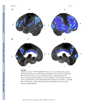
FIGURE 3.
18
Alzheimer’s disease: F-FDG brain PET/CT study of a 81-year-old man with cognitive
decline during the past few months. The left and right lateral (A) and left and right medial
(B) 3-dimensional stereotactic surface projection images of FDG hypometabolism
demonstrate glucose hypometabolism involving the bilateral frontal, temporal, and parietal
cortices (compared with a normal brain database, Mim software 6.2. Light blue, −1 SD; dark
blue, −2 SDs; purple, −3SDs), with sparing of the sensorimotor and occipital cortices,
features consistent with AD.
NIH-PA Author Manuscript
Clin Nucl Med. Author manuscript; available in PMC 2015 February 18.