Page 17 - HBC Booklet - 2019
P. 17

Eur Spine J (2011) 20:1791-1795                                                                                                                                                                                          1793


                        harvested. To harvest the cells, cultures were rinsed in PBS                                                          a                                                                •

                        followed by osteoblast detachment using  0.25% Trypsin­


                        EDT A  (lnvitrogen/Carlsbad,  CA,  USA). Trypsin  incuba­

                        tion was stopped by adding culture medium t o   a 1: 1 ratio,


                        and  cell suspensions were washed  twice  in PBS  prior  to


                        cell  viability  and  cell  count  determination.  Finally,  cell

                        yield per gram bone tissue was calculated.


                        Evaluation of population doubling times


                        and cell viability


                        Cells  harvested  after  a 3 weeks  observation  period  were

                                                           2
                        seeded into 7 5   cm tissue culture flasks.  After confluence

                        was  reached,  cells  were  harvested  and  counted  as previ­


                        ously described. Population doubling times were calculated                                                             b                                                                                          •

                        and cell viability was determined using the trypan blue dye

                        exclusion test.


                        Statistical evaluation


                        SPSS 14.0 was used for statistical evaluation. Groups were

                        compared using a 2-tailed student's t test at a 0.05 level of                                                                                                                                                           I


                        significancy.


                        Results


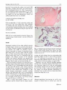
                        Histological  evaluation of bone chips collected  using  the

                        kerrington rongeur showed-as expected-intact tiny pie­


                        ces  of bone.  Concentric lamellae  surrounding the Haver­                                                          Fig. 1  a Laminectomy bone chips demonstrating intact lamellar bone

                        sian  Channels  were  visible  and  osteoblasts  could  be                                                          structure,  osteoblasts  within  their  lacunae  and blood vessel supply.


                        observed  surrounded  by  lacunae  of  extracellular  matrix.                                                       b  Bone  shavings  showing  disrupted  bone  structure  with  single
                                                                                                                                            osteoblasts (arrol-v)  deprived from their extracellular  matrix. Blood
                        Intact blood  vessels were also visible  within the samples                                                         vessels  are not observed


                        (Fig.  la).  Slides  obtained  from  bone  shavings  showed  a

                        different  aspect:  Bone  tissue  was  disintegrated  into  tiny                                                    Average  cell  yield  obtained  from  the corresponding high


                        fragments,  and  although  remnants  of  the  interstitial                                                          speed burr samples was about 7x lower with an average of

                                                                                                                                                               5
                        lamellae  were  observed, osteoblasts  were  separated  from                                                        1.73  x 10 osteoblasts  per  gram bone  (p < 0.01,  Fig. 3).

                        their  lacunae  and  located  loosely  between  the  lamellar                                                       Despite the highly different emigration time span and cell

                        fragments. Blood vessels were not observed (Fig.  lb).                                                              yield, viability in both study groups  was equal at 98%.


                             Regarding osteoblast emigration, tissue harvested by the                                                            Differences  were  also  observed  regarding  population


                        kerrington rongeur demonstrated reliable osteoblast release                                                         doubling times:  osteoblasts emigrated  from rongeur bone

                        after average 5.6 days. Corresponding bone tissue obtained                                                          chips  duplicated  within  50.5 h  while  the  corresponding


                        via  high  speed  drill  showed  a  high  variation  regarding                                                      bone  shavings  required  121 h  for  cell  cycle  completion

                        osteoblast delivery: Although all rongeur samples from all                                                          (p < 0.01; Fig. 4). After 3 weeks o f   in vitro culture, posi­


                        patients-no  matter whether  obese,  osteoporotic  or  after                                                        tive  Alizarin  red  staining  indicating  mineralization  of

                        cortisol  treatment--demonstrated  successful  osteoblast                                                           monolayer tissue was only visible i n   cultures derived from


                        delivery to the culture dish, only 8 out of 14 corresponding                                                        rongeur bone chips (Fig. 5a, b ).

                        samples (57%) harvested via high speed burr were able to


                        do so (p < 0.024). The time span of osteoblast delivery-if


                        any-was highly variable between 7 and 30 days (average                                                              Discussion

                        time span 14.8 days; p < 0.003; Fig. 2).


                             After  a  3 weeks  culture  period,  average  1.25 x 10                                               6        Although  larninectomy  bone  shavings  are  used  by  spine

                        osteoblasts  could  be  obtained  from  the  rongeur samples.                                                       surgeons  to  enhance  fusion  in  cervical  spine,  only  one


                                                                                                                                                                                                                                      � Springer
   12   13   14   15   16   17   18   19   20