
Page 9 - HBC Booklet - 2019
P. 9
116 International Orthopaedics (SICOT) (2008) 32:115-119
t h e principle of adequate decompression, repositioning, Table 1 Preoperative radiological features o f 136 patients
fusion and adequate fixation in order t o achieve a good Level of instability Number of patients
outcome [3]. These operative procedures can be performed
via either an anterior or posterior approach, with a choice of L. 111-V 46
autogenous graft and allograft can be used for fusion. L. I V -V 9 0
Usually a n autogenous graft is the most commonly preferred, Grade of the slip
because it provides a much better outcome, and the most Meyerding Grade 1 105
popular donor site is the iliac crest bone. However, many Meyerding Grade 2 31
studies have shown that the iliac crest autogenous g raft
harvest is not risk-free, reporting an overall complication rate ment; (3) progressive radiculopathy; ( 4) radiologically
ranging from 9.4% to 49% [2]. We have undertaken this proven instability. An informed consent was obtained prior
retrospective study to assess the outcome of posterolateral t o operation. Spinal fusion was then assessed by plain
fusion (PLF) using laminectomy bone chips for the treatment lumbar spine radiographs at 4, 8, and 24 months after
o f lumbar spondylolisthesis. operation. Additional plain lumbar spine radiographs were
performed on 60 patients showing solid fusion mass after
removal of the spinal implants.
Materials and methods
From January 1993 t o December 2003, a total of 136 Results
patients (98 females and 38 males; aged 16-76 years, with
a n average of 46 years) diagnosed with lumbar spondylolis All patients underwent the procedure smoothly, with
thesis (LS) by plain lumbar radiographs, treated and average operative time for one-level lesions being 1 h and
followed-up well at our Orthopaedic Division were includ 30 min, while 2 h and 15 min was spent for two-level
ed in this study. All patients presented with persistent low lesions. Blood transfusion was routinely given for patients
back pain with radiculopathy and intermittent claudication. with two-level lesions and only to one-level lesion patients
Computed tomography (CT) scans or magnetic resonance whose preoperative haemoglobin levels were less than 1 1 g/
imagings (MRis) were performed in all patients to identify dl. One-hundred and twenty-nine cases (94.85%) developed
other associated lesions, such as ruptured disc and spinal solid fusion mass at 8 months post-operation (Figs. 2, 3)
stenosis. Each and every patient underwent a near total with failed fusion noted in seven cases (5.15%). Fusion rate
posterior decompression laminectomy with foraminotomy for the one-level lesion group was 93% (84/90) and 97%
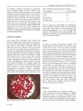
and PLF with laminectomy bone chips (Fig. 1) as bone (45/46) for the two-level lesion group. Fusion was assessed
graft, followed by reduction of the slipped vertebra with based on the criteria summarised in Table 2. No mortality
transpedicle screws and the A-0 or Trifix Reduction spinal nor morbidity was encountered in our series and our
system implants. Tue main preoperative radiographic patients were discharged on the fifth post-operative day
characteristics are summarised in Table 1. All patients with a Knight-Taylor body brace. All o f the 129 patients
fulfilled the following criteria: (1) intractable low-back pain that developed solid fusion after the operation claimed t o
and/or sciatic pain; (2) failed previous conservative treat- have relief of symptoms (low-back pain with radiculopathy
and intermittent claudication) noted immediately after the
procedure, and throughout the follow-up period. Sixty
patients returned for removal of the spinal implants four
years after operation, repeat plain lumbar radiographs
(Fig. 4) after removal of implants showed solid fusion
mass between the involved vertebrae.
Discussion
Lumbar spondylolisthesis was originally described as a
cause of obstruction in labour by Herbiniaux, a Belgian
obstetrician in 1782 [I, 6, 7], and its was Rokitansky who i s
credited for describing it a s a pathological entity [1]. Since
then, many authors have dedicated their time and effort t o
Fig. 1 Laminectomy bone-chip graft th e study and search for the aetiology of spondylolisthesis.
� Springer