
Page 13 - Current techniques in canine and feline neurosurgery_2017_Neat
P. 13
4 Section I: Diagnostics and Planning
B
Camera head
A Telescope
C
Telescope holder
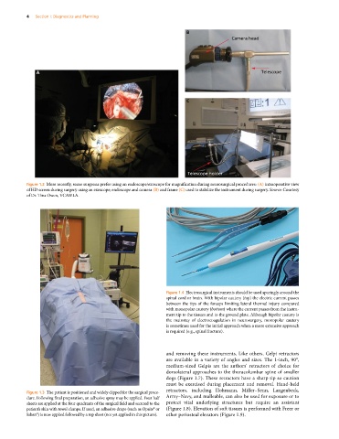
Figure 1.2 More recently, some surgeons prefer using an endoscope/exoscope for magnification during neurosurgical procedures: (A) intraoperative view
of HD screen during surgery using an exoscope; endoscope and camera (B) and frame (C) used to stabilize the instrument during surgery. Source: Courtesy
of Dr. Tina Owen, VCAWLA.
Figure 1.4 Electrosurgical instruments should be used sparingly around the
spinal cord or brain. With bipolar cautery (top) the electric current passes
between the tips of the forceps limiting lateral thermal injury compared
with monopolar cautery (bottom) where the current passes from the instru-
ment tip to the tissues and to the ground plate. Although bipolar cautery is
the mainstay of electrocoagulation in neurosurgery, monopolar cautery
is sometimes used for the initial approach when a more extensive approach
is required (e.g., spinal fracture).
and removing these instruments. Like others, Gelpi retractors
are available in a variety of angles and sizes. The 1‐inch, 90°,
medium‐sized Gelpis are the authors’ retractors of choice for
dorsolateral approaches to the thoracolumbar spine of smaller
dogs (Figure 1.7). These retractors have a sharp tip so caution
must be exercised during placement and removal. Hand‐held
Figure 1.3 The patient is positioned and widely clipped for the surgical proce- retractors, including Hohmann, Miller–Senn, Langenbeck,
dure. Following final preparation, an adhesive spray may be applied. Four half Army–Navy, and malleable, can also be used for exposure or to
sheets are applied at the four quadrants of the surgical field and secured to the protect vital underlying structures but require an assistant
patient’s skin with towel clamps. If used, an adhesive drape (such as Opsite® or (Figure 1.8). Elevation of soft tissues is performed with Freer or
Ioban®) is now applied followed by a top sheet (not yet applied in this picture). other periosteal elevators (Figure 1.9).