
Page 38 - Current techniques in canine and feline neurosurgery_2017_Neat
P. 38
30 Section I: Diagnostics and Planning
A B
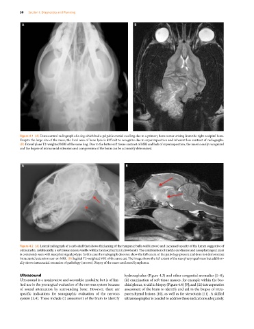
Figure 4.1 (A) Dorsoventral radiograph of a dog which had a palpable cranial swelling due to a primary bone tumor arising from the right occipital bone.
Despite the large size of the mass, the focal area of bone lysis is difficult to recognize due to superimposition and inherent low contrast of radiographs.
(B) Dorsal plane T2‐weighted MRI of the same dog. Due to the better soft tissue contrast of MRI and lack of superimposition, the mass is easily recognized
and the degree of intracranial extension and compression of the brain can be accurately determined.
A B
Figure 4.2 (A) Lateral radiograph of a cat’s skull that shows thickening of the tympanic bulla wall (arrow) and increased opacity of the lumen suggestive of
otitis media. Additionally, a soft tissue mass is visible within the nasopharynx (arrowhead). The combination of middle ear disease and nasopharyngeal mass
is commonly seen with nasopharyngeal polyps. In this case the radiograph does not show the full extent of the pathology present and does not demonstrate
intracranial extension seen on MRI. (B) Sagittal T2‐weighted MRI of the same cat. The image shows the full extent of the nasopharyngeal mass but addition-
ally shows intracranial extension of pathology (arrows). Biopsy of the mass confirmed lymphoma.
Ultrasound hydrocephalus (Figure 4.3) and other congenital anomalies [5–8];
Ultrasound is a noninvasive and accessible modality, but is of lim- (ii) examination of soft tissue masses, for example within the bra-
ited use in the presurgical evaluation of the nervous system because chial plexus, to aid in biopsy (Figure 4.4) [9]; and (iii) intraoperative
of sound attenuation by surrounding bone. However, there are assessment of the brain to identify and aid in the biopsy of intra-
specific indications for sonographic evaluation of the nervous parenchymal lesions [10], as well as for stereotaxis [11]. A skilled
system [3,4]. These include (i) assessment of the brain to identify ultrasonographer is needed to address these indications adequately.