
Page 67 - Current techniques in canine and feline neurosurgery_2017_Neat
P. 67
Chapter 6: Minimum Database: Spinal Surgery 61
Size Alterations
• Microcytosis can be caused by congenital portosystemic shunt or
iron deficiency, or be a normal finding in some canine breeds
(Shiba Inu, Akita, Chow Chow).
• Macrocytosis can be the result of regenerative anemias or myelo-
proliferative diseases.
Anemia
• Regenerative: with evidence of bone marrow response to a
decreased hematocrit, that typically is based on the presence of
reticulocytes in circulation. Associated with acute blood loss or
hemolysis (immune‐mediated hemolytic anemia, toxicity, eryth-
roparasites, hereditary diseases).
• Nonregenerative: with no evidence of bone marrow response,
usually normocytic/normochromic. Associated with underlying
conditions that affect bone marrow erythrocyte production such
as neoplasia (leukemia or metastatic), aplastic anemia, infectious
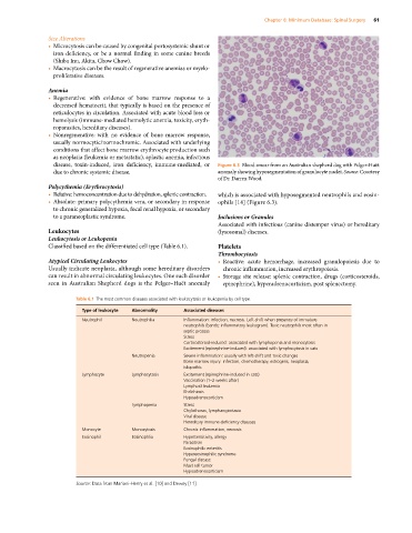
disease, toxin‐induced, iron deficiency, immune‐mediated, or Figure 6.3 Blood smear from an Australian shepherd dog with Pelger‐Huët
due to chronic systemic disease. anomaly showing hyposegmentation of granulocyte nuclei. Source: Courtesy
of Dr. Darren Wood.
Polycythemia (Erythrocytosis)
• Relative: hemoconcentration due to dehydration, splenic contraction. which is associated with hyposegmented neutrophils and eosin-
• Absolute: primary polycythemia vera, or secondary in response ophils [14] (Figure 6.3).
to chronic generalized hypoxia, focal renal hypoxia, or secondary
to a paraneoplastic syndrome. Inclusions or Granules
Associated with infectious (canine distemper virus) or hereditary
Leukocytes (lysosomal) diseases.
Leukocytosis or Leukopenia
Classified based on the differentiated cell type (Table 6.1). Platelets
Thrombocytosis
Atypical Circulating Leukocytes • Reactive: acute hemorrhage, increased granulopoiesis due to
Usually indicate neoplasia, although some hereditary disorders chronic inflammation, increased erythropoiesis.
can result in abnormal circulating leukocytes. One such disorder • Storage site release: splenic contraction, drugs (corticosteroids,
seen in Australian Shepherd dogs is the Pelger–Huët anomaly epinephrine), hyperadrenocorticism, post splenectomy.
Table 6.1 The most common diseases associated with leukocytosis or leukopenia by cell type.
Type of leukocyte Abnormality Associated diseases
Neutrophil Neutrophilia Inflammation: infection, necrosis. Left‐shift when presence of immature
neutrophils (bands; inflammatory leukogram). Toxic neutrophils most often in
septic process
Stress
Corticosteroid‐induced: associated with lymphopenia and monocytosis
Excitement (epinephrine‐induced): associated with lymphocytosis in cats
Neutropenia Severe inflammation: usually with left‐shift and toxic changes
Bone marrow injury: infection, chemotherapy, estrogens, neoplasia,
idiopathic
Lymphocyte Lymphocytosis Excitement (epinephrine‐induced in cats)
Vaccination (1–2 weeks after)
Lymphoid leukemia
Ehrlichiosis
Hypoadrenocorticism
Lymphopenia Stress
Chylothorax, lymphangiectasia
Viral disease
Hereditary immune‐deficiency diseases
Monocyte Monocytosis Chronic inflammation, necrosis
Eosinophil Eosinophlia Hypersensitivity, allergy
Parasitism
Eosinophilic enteritis
Hypereosinophilic syndrome
Fungal disease
Mast cell tumor
Hypoadrenocorticism
Source: Data from Marioni‐Henry et al. [10] and Dewey [11].