
Page 85 - Current techniques in canine and feline neurosurgery_2017_Neat
P. 85
80 Section I: Diagnostics and Planning
A and CT myelography were equally sensitive in the diagnosis of
intervertebral disc herniation [53].
MRI is equivalent or superior to myelography and CT for
localizing spinal cord compression due to intervertebral disc her
niation [42,54,55]. MRI also facilitates assessment of the spinal cord
parenchyma for edema and myelomalacia which can be prognostic
indicators. Spinal cord edema is characterized by T2 hyperintensity
and swelling of the spinal cord. Hemorrhagic myelomalacia can
also be seen as a decreased signal on gradient echo (T2*) images [56].
Although spinal cord lesions can provide insight into prognosis, the
degree of spinal cord compression is not correlated with neurologi
cal status at presentation or the outcome [57]. Heavily T2‐weighted
images demonstrate the subarachnoid space similarly to myelogra
B
phy and can be useful in determining the site and severity of acute
spinal cord compression as length of spinal cord compression has
been associated with lower odds of returning to ambulation [58,59].
Contrast enhancement can occur in intervertebral disc herniation
due to focal meningitis and formation of granulation tissue but is
not related to clinical signs or pathological features [60,61].
Lumbosacral Disease
Many of the signs of lumbosacral disease are the same as seen with
intervertebral disc disease. Myelography is limited in evaluating
lumbosacral disease due to the variability in termination of the
dural sac (between L6 and S1). Approximately 20% of dogs have a
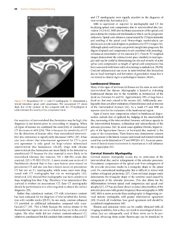
Figure 7.13 Noncontrast CT (A) and CT‐myelography (B) demonstrate a dural sac that ends cranial to the sacrum [62]. Additionally, mye
ventral extradural spinal cord compression. The noncontrast CT shows lography does not allow evaluation of lateral lesions such as stenosis
slight loss of the epidural fat but compared with the CT‐myelogram it of the intervertebral foramen [63]. As a result CT and MRI are
underestimates the severity of the compressive lesion. superior tests for the evaluation of the lumbosacral region.
Signs of nerve root compression related to lumbosacral degen
eration include loss of epidural fat, bulging of the intervertebral
the suspicion of intervertebral disc herniation may be high, this disc, narrowing of the intervertebral foramen, soft tissue opacity in
diagnosis is not known prior to proceeding to imaging. When the intervertebral foramen, subluxation and osteoarthrosis of the
all types of lesions are combined the sensitivity of unenhanced articular processes [64]. In most cases epidural fibrosis, hypertro
CT decreases to 66% [26]. This is because the sensitivity of CT phy of the ligamentum flavum, or herniated disc material is the
for the detection of lesions other than mineralized interverte cause of the compression. These lesions may demonstrate contrast
bral disc extrusions is significantly decreased (40%) [29]. It has enhancement in the lateral recesses and dorsal and ventral vertebral
also been shown that interobserver agreement for CT is poor canal that can be detected on CT and MRI [65–67]. Accurate assess
and agreement is only good for large‐volume mineralized ment of lateral recess involvement is important as it will influence
intervertebral disc herniations [26,29]. Dogs with chronic the surgical plan [65].
intervertebral disc herniation are more likely to be detected on
unenhanced CT because the disc material is more likely to be Cervical Stenotic Myelopathy
mineralized (chronic disc material, 745 ± 288 HU; acute disc Cervical stenotic myelopathy occurs due to protrusion of the
material, 219 ± 95 HU) [31,51]. A more recent case series of 11 intervertebral disc and/or enlargement of the articular processes.
Dachshunds showed that in four cases lesions were missed on Dorsolateral compression of the spinal cord from enlargement of
precontrast CT and one case had a lesion noted on precontrast the articular processes results in a triangular shape to the spinal
CT but not on CT myelography, while some cases were diag cord. On conventional myelography this is difficult to detect with
nosed with CT myelography but not on myelography [32]. routine orthogonal projections [27]. Cross‐sectional images easily
Israel et al. [31] showed that myelography was more sensitive in demonstrate the triangular shape of the vertebral canal caused by
dogs weighing less than 5 kg. Therefore, if no mineralized disc enlargement of the articular processes. This also allows for the
material is evident on noncontrast CT, then CT myelography differentiation between spinal cord compression and spinal cord
should be performed as it is often required to obtain the correct atrophy [27]. CT has also been shown to detect abnormalities of the
diagnosis [29]. articular processes with greater frequency than radiography or MRI
Rather than intrathecal contrast, CT with intravenous contrast [68]. MRI is more accurate than myelography in diagnosing cer
has been evaluated for the diagnosis of intervertebral disc hernia vical stenotic myelopathy, with greater interobserver agreement
tion with variable results [29,53]. In one study, contrast‐enhanced [69]. Overall, all modalities have good agreement and should be
CT provided no additional information compared with unen considered complementary [68].
hanced CT [29]. This is likely because the venous sinuses in the Flexion and extension views can be readily obtained with all
thoracolumbar region are not as evident as they are in the cervical imaging modalities, but as they can result in neurological deterio
region. The other study did not evaluate contrast‐enhanced CT ration they are infrequently used. If these views are to be per
relative to unenhanced but did conclude that contrast‐enhanced CT formed, obtaining them under fluoroscopy can be beneficial by