
Page 1078 - Veterinary Toxicology, Basic and Clinical Principles, 3rd Edition
P. 1078
1010 SECTION | XV Mycotoxins
VetBooks.ir TABLE 71.1 (Continued)
Number of
Animals Dose & Route Duration Toxic Effects Reference
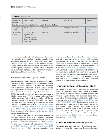
Reported Fumonisin Concentrations From Naturally-Occurring Outbreaks
34 pigs from 105 155 ppm FB 1 Unknown Lethal pulmonary edema Harrison et al.
2 farms (1990)
16 pigs from All feed samples Unknown Lethal pulmonary edema Osweiler et al.
9 farms associated with (1992)
pulmonary edema
contained $20 ppm
FB 1 to a maximum of
330 ppm FB 1
In ultrastructural studies using immersion fixed lungs, observed as early as 2 days after the initiation of treat-
the endothelium was found to be swollen, vacuolated, and ment with a lethal dose (Gumprecht et al., 1998), and at a
sometimes missing in pigs with pulmonary edema concentration as low as 23 ppm when fed for 14 days
(Haschek et al., 1992). Additional studies using intravas- (Motelin et al., 1994). Long-term fumonisin exposure can
cularly perfused lungs (to allow better examination of the result in fibrosis or development of hyperplastic nodules
vascular system) demonstrated accumulations of fragmen- in the liver (Casteel et al., 1993; Harrison et al., 1990).
ted membranous material in the cytocavitary region of Ultrastructurally, large accumulations of proteinaceous
endothelial cells (Gumprecht et al., 1998). and membranous material were observed in the space of
Disse in pigs that developed fumonisin-induced pulmo-
nary edema (Haschek et al., 1992). Hepatocytes lost
Fumonisins in Swine-Hepatic Effects microvilli from their sinusoidal face while numerous
Kupffer cells contained multilamellar bodies.
Hepatic changes in pigs exposed to fumonisins include
elevation of liver associated enzyme activities, altered
clinical chemistries, changes in sphingolipid parameters, Fumonisins in Swine-Cardiovascular Effects
and morphological alterations. In pigs, hepatic toxicity
occurs prior to the development of pulmonary edema, and Fumonisins have been shown to decrease left ventricular
alterations are time and dose-dependent (Motelin et al., contractility, heart rate, cardiac output, mean arterial pres-
1994). Increased activities of serum enzymes such as sure, arterial and mixed venous blood O 2 tensions, and
alkaline phosphatase (ALP), aspartate aminotransferase systemic oxygen delivery, while increasing mean pulmo-
(AST) and gamma glutamyl transpeptidase (GGT) and nary artery pressure, oxygen consumption, and oxygen
concentrations of total bilirubin, bile acids, and choles- extraction ratio in swine (Constable et al., 2000; Smith
terol have been reported as early as 1 day after the initia- et al., 1996a,b, 1999, 2000). The decrease in cardiac con-
tion of fumonisin exposure (Colvin et al., 1993; tractility leads to acute left ventricular failure and pulmo-
Gumprecht et al., 1998; Harrison et al., 1990; Haschek nary edema in pigs exposed to high concentrations of
et al., 1992; Motelin et al., 1994; Osweiler et al., 1992). fumonisin in feed. Chronic exposure to lower levels of
These alterations reflect hepatocyte damage as well as fumonisin leads to the development of right ventricular
altered hepatic function. hypertrophy and medial hypertrophy of the small pulmo-
Morphologic alterations are dose related and progres- nary arteries in pigs, likely a result of pulmonary hyper-
sive with continued ingestion of fumonisins. Following tension (Casteel et al., 1994).
short term exposure, changes include hepatic cord disor-
ganization, cytoplasmic vacuolation, apoptosis, scattered Fumonisins in Swine-Immunologic Effects
necrosis, and increased cell proliferation (Gumprecht
et al., 1998; Harrison et al., 1990; Motelin et al., 1994; Fumonisins have also been shown to predispose pigs to
Osweiler et al., 1992). Histologic alterations were respiratory disease. In one case-control study, swine farms