
Page 179 - Live-cellanalysis handbook
P. 179
IncuCyte® Fluorescence Neurite Analysis Assay
Protocol Overview: IncuCyte Annexin V NIR Reagent
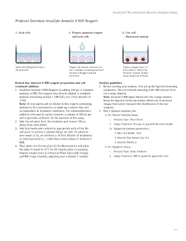
1. Seed cells 2. Prepare apoptosis reagent 3. Live-cell
and treat cells fluorescent analysis
Seed cells (100 μL/well) into a Prepare the desired treatments at Capture images every 2-3
96-well plate. 2X in medium containing IncuCyte hours (20X or 10X) in the
®
Annexin V Reagent and add IncuCyte System. Analyze
®
treatment. using integrated software.
Desired day: Annexin V NIR reagent preparation and cell Analysis guidelines
treatment addition 1. Before starting your analysis, first set up the Spectral Unmixing
1. Solubilize Annexin V NIR Reagent by adding 100 μL of complete parameters. We recommend removing 2-4% NIR channel from
medium or PBS. The reagent may then be diluted in complete the orange channel.
medium containing at least 1 mM CaCl 2 for a final dilution of Note: Annexin V NIR signal bleeds into the orange channel.
1:100. Using the Spectral Unmixing feature allows you to produce
Note: All test agents will be diluted in this reagent-containing images that better represent the distribution of the two
medium to 2X concentration, so make up a volume that will reagents.
accommodate all treatment conditions. The volumes/dilutions 2. Run 2 separate analysis jobs.
added to cells may be varied; however, a volume of 200 μL per a. For Neurite Analysis Assay:
well is generally sufficient for the duration of the assay. i. Analysis Type: NeuroTrack
2. Take the cell plate from the incubator and remove 100 μL
media from wells (50%). ii. Image Channels: Orange to quantify Neurite Length
3. Add treatments and controls to appropriate wells of the 96- iii. Suggested analysis parameters:
well plate to achieve a volume 200 μL per well. As solutions 1. Min Cell Width: 15.0
were made at 2X, we will have a 1X final dilution of treatments
in individual wells (i.e., 1:200 final concentration of Annexin V 2. Neurite Fine Sensitivity: 0.5
NIR). 3. Neurite Width: 2
4. Place plate into the IncuCyte S3 for Neuroscience and allow b. For Apoptosis Assay:
the plate to warm to 37˚C for 30 minutes prior to scanning.
Acquire images every 2-3 hours in Phase (optional), Orange, i. Analysis Type: Basic Analyzer
and NIR image channels, adjusting scan schedule if needed. ii. Image Channels: NIR to quantify apoptotic cells
177