
Page 119 - Differential Diagnosis in Small Animal Cytology, The Skin and Subcutis
P. 119
er 8
Chapt
106
VetBooks.ir
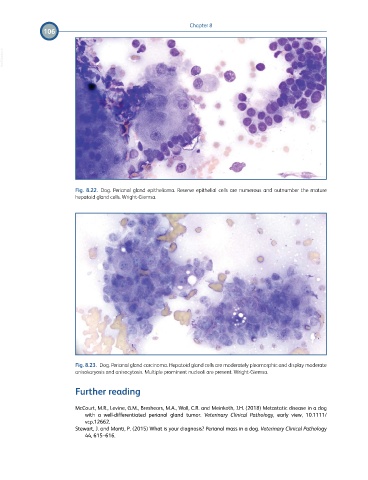
Fig. 8.22. Dog. Perianal gland epithelioma. Reserve epithelial cells are numerous and outnumber the mature
hepatoid gland cells. Wright-Giemsa.
Fig. 8.23. Dog. Perianal gland carcinoma. Hepatoid gland cells are moderately pleomorphic and display moderate
anisokaryosis and anisocytosis. Multiple prominent nucleoli are present. Wright-Giemsa.
Further reading
McCourt, M.R., Levine, G.M., Breshears, M.A., Wall, C.R. and Meinkoth, J.H. (2018) Metastatic disease in a dog
with a well-differentiated perianal gland tumor. Veterinary Clinical Pathology, early view, 10.1111/
vcp.12662.
Stewart, J. and Monti, P. (2015) What is your diagnosis? Perianal mass in a dog. Veterinary Clinical Pathology
44, 615–616.