
Page 146 - Differential Diagnosis in Small Animal Cytology, The Skin and Subcutis
P. 146
Mesenchymal Tumours and Other Neoplasms
133
VetBooks.ir
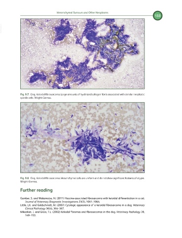
Fig. 9.7. Dog. Keloidal fibrosarcoma. Large amounts of hyalinized collagen fibrils associated with slender neoplastic
spindle cells. Wright-Giemsa.
Fig. 9.8. Dog. Keloidal fibrosarcoma. Mesenchymal cells are uniform and do not show significant features of atypia.
Wright-Giemsa.
Further reading
Gumber, S. and Wakamatsu, N. (2011) Vaccine-associated fibrosarcoma with keloidal differentiation in a cat.
Journal of Veterinary Diagnostic Investigations 23(5), 1061–1064.
Little, L.K. and Goldschmidt, M. (2007) Cytologic appearance of a keloidal fibrosarcoma in a dog. Veterinary
Clinical Pathology 36(4), 364–367.
Mikaelian, I. and Gross, T.L. (2002) Keloidal fibromas and fibrosarcomas in the dog. Veterinary Pathology 39,
149–153.