
Page 185 - Differential Diagnosis in Small Animal Cytology, The Skin and Subcutis
P. 185
Chapt er 10
172
VetBooks.ir
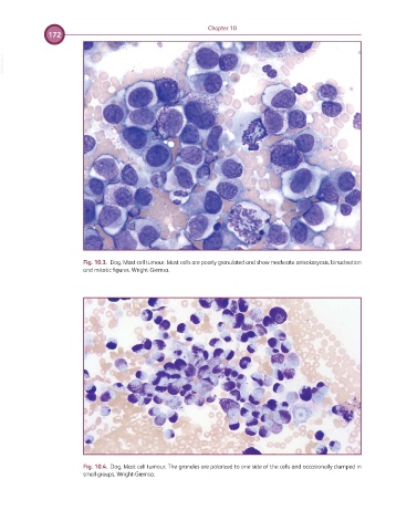
Fig. 10.3. Dog. Mast cell tumour. Mast cells are poorly granulated and show moderate anisokaryosis, binucleation
and mitotic figures. Wright-Giemsa.
Fig. 10.4. Dog. Mast cell tumour. The granules are polarized to one side of the cells and occasionally clumped in
small groups. Wright-Giemsa.