
Page 60 - Differential Diagnosis in Small Animal Cytology, The Skin and Subcutis
P. 60
Inflammatory Lesions
47
VetBooks.ir Pearls and Pitfalls
Idiopathic sterile nodular panniculitis is a descriptive term used to indicate a sterile
•
inflammatory disease of the subcutaneous fat for which the triggering aetiology is
unknown. It presents as multiple subcutaneous nodules, often ulcerated, fistulated and
draining a lipid material mixed with blood. The trunk is the most commonly affected
anatomical area. This form is often associated with systemic clinical signs, such as fever,
anorexia and malaise. The Dachshund breed is considered at increased risk.
• Juvenile sterile granulomatous dermatitis and lymphadenitis (also called puppy strangles
or juvenile cellulitis, as the inflammation often extends to the subcutis) is a disease of
unknown origin affecting young dogs (puppies). Clinically, it is characterized by
swelling and exudation of the skin of ears, eyelids, lips, nose and mucucutaneous
junctions. Submandibular lymph nodes may be enlarged and systemic clinical signs
of malaise are common. Cocker Spaniel, Dachshund and Gordon Setter dogs seem
more frequently represented. A pyogranulomatous process with prevalence of neu-
trophils and macrophages is observed on cytology.
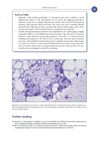
Fig. 6.15. Dog. Panniculitis secondary to trauma. The background is proteinaceous and contains numerous punc-
tate fat droplets. Multiple macrophages are seen. They have abundant cytoplasm containing high numbers of lipid
vacuoles. Wright-Giemsa.
Further reading
Countreary, C.L., Outerbridge, C.A., Affolter, V., Kass, P.H. and White, S.D. (2015) Canine sterile nodular pannicu-
litis: a retrospective study of 39 dogs. Veterinary Dermatology 26, 451–458.
O’Kell, A.L., Inteeworn, N., Diaz, S.F., Saunders, G.K. and Panciera, D.L. (2010) Canine sterile nodular panniculitis:
a retrospective study of 14 cases. Journal of Veterinary Internal Medicine 24, 278–284.