
Page 1000 - Equine Clinical Medicine, Surgery and Reproduction, 2nd Edition
P. 1000
Cardiovascular system 975
VetBooks.ir 8.12
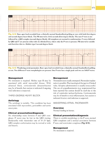
Fig. 8.12 Base–apex lead recorded from a clinically normal Standardbred gelding at rest, with both first-degree
and second-degree heart block. The PR interval is >0.42 seconds (first-degree block). The next P wave is not
followed by a QRS complex (second-degree block). All complexes are normal in conformation. P waves followed
by QRS and T are present either side of a blocked beat, where only the P wave is present. PR interval is variable
and therefore this is a Mobitz type I second-degree block.
8.13
Fig. 8.13 Wandering atrial pacemaker. Base–apex lead recorded from a clinically normal Standardbred gelding
at rest. Two different P wave morphologies are present. One P wave has a single peak and two are bifid P waves.
Management Management
No treatment is required. Mobitz type II may be Treatment is not usually attempted. Pacemaker implan-
associated with atrial myocardial disease. With tation is possible. Pharmacological therapy is of limited
advanced block, corticosteroids (dexamethasone) value. Atropine administration is usually unsuccessful.
may be of benefit, but caution is indicated if ongoing The use of sympathomimetics (e.g. isoproterenol) has
viral infection is suspected. been reported but caution should be used due to the
risk of ventricular tachyarrhythmias. Corticosteroids
THIRD-DEGREE HEART BLOCK may be of benefit to treat inflammation, but caution is
indicated if active viral infection is suspected.
Aetiology
The aetiology is variable. This condition has been WANDERING ATRIAL PACEMAKER
associated with myocarditis, pericarditis and aortic
aneurysms. Overview
Normal finding in horses.
Clinical presentation/diagnosis
No relationship exists between P and QRS com- Clinical presentation/diagnosis
plexes. P waves may be lost in the QRS tracing. There is variable morphology to the P wave, normal
Bradycardia with ventricular rate of 10–20 bpm. QRS complexes and a normal association between
QRS complex conformation may be normal or wide P wave and ventricular complexes (Fig. 8.13).
and bizarre.
Management
No treatment is required.