
Page 1017 - Equine Clinical Medicine, Surgery and Reproduction, 2nd Edition
P. 1017
992 CHAPTER 8
VetBooks.ir in platelet aggregation and fibrin deposition on the result in murmurs due to higher pressure differences
Prior injury, or that initiated by bacteria, results
across the valves than on the right side. Mural lesions
endocardial surface. This results in the formation of
vegetative lesions consisting of platelets, bacteria and are unlikely to be associated with a murmur. Cardiac
arrhythmia secondary to bacterial emboli to the
fibrin at the site of infection (Fig. 8.26). Damage to myocardium has been reported. With extensive dam-
valvular endocardium results in insufficiency and, age to valves, valvular insufficiency may lead to heart
when extensive, can precipitate cardiac failure. failure. Clinical signs may also be associated with the
Vegetative lesions are often friable and thromboem- sequelae of endocarditis, such as renal infarction.
boli might develop. Thromboemboli from the aortic
or left AV valve may cause obstruction of vital ves- Differential diagnosis
sels such as those supplying the kidneys, the brain Parasitic endocarditis (uncommon, aortic valve),
or even the heart itself. Immune-complex deposition congenital heart disease, acquired valve insuf-
may also be associated with systemic disease such as ficiency, abscessation, neoplasia, septicaemia and
polyarthritis. polyarthritis should be considered.
Clinical presentation Diagnosis
A common presentation in the horse is fever, which is Clinical signs are often unremarkable; however,
often intermittent. Tachypnoea, tachycardia, weight sudden onset of a murmur associated with pyrexia
loss, anorexia and depression are also common. should raise concern. A complete blood count and
A variable lameness may also be present. Cardiac blood culture are valuable tools in the diagnosis
murmurs are not always present with endocarditis, of endocarditis. Leucocytosis with neutrophilia is
but suspicion should be raised when a new-onset common. Non-regenerative anaemia consistent with
murmur is associated with pyrexia and ill-thrift. anaemia of chronic disease may also be present.
Lesions on the left side of the heart are more likely to Hyperfibrinogenaemia is also common. Blood cul-
ture may be unrewarding, often because of previous
antimicrobial administration and low levels of circu-
lating microbes, but should be performed. Serial blood
8.26 cultures may be of benefit (e.g. every 2 hours for three
cultures). Collection of blood culture during periods
of pyrexia or immediately prior to a febrile period
may be more useful. The organisms most commonly
identified in endocarditis in the horse are Streptococcus
zooepidemicus, Actinobacillus equuli and staphylococci.
E. coli has also been identified.
Echocardiography is the most useful tool in the
diagnosis of endocarditis. Valvular deformity and
vegetative lesions are relatively easy to visualise. Non-
valvular endocarditis is more difficult to identify.
Usually there are no radiographic abnormalities.
Arrhythmias are possible with endocarditis. Premature
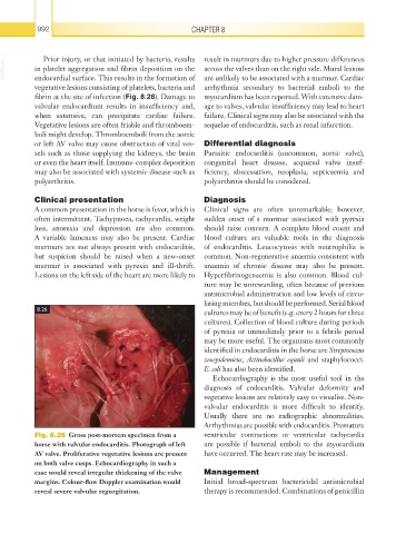
Fig. 8.26 Gross post-mortem specimen from a ventricular contractions or ventricular tachycardia
horse with valvular endocarditis. Photograph of left are possible if bacterial emboli to the myocardium
AV valve. Proliferative vegetative lesions are present have occurred. The heart rate may be increased.
on both valve cusps. Echocardiography in such a
case would reveal irregular thickening of the valve Management
margins. Colour-flow Doppler examination would Initial broad-spectrum bactericidal antimicrobial
reveal severe valvular regurgitation. therapy is recommended. Combinations of penicillin