
Page 1079 - Equine Clinical Medicine, Surgery and Reproduction, 2nd Edition
P. 1079
1054 CHAPTER 10
VetBooks.ir unsafe or likely to injure themselves or others. The Thoracolumbar region (T3–L3)
Severe hindlimb ataxia and/or spasticity with normal
author tends to perform this blindfolded part of the
gait assessment in a ménage with plenty of space and
spinal cord lesion. There is likely to be normal tail
a soft landing, rather than on a hard trot-up surface, forelimbs is usually indicative of a thoracolumbar
in a clinic or the horse’s stable. and anal tone, with no neurogenic atrophy. There
may be hypoalgesia or analgesia caudal to the cranial
Grading gait abnormalities border of the lesion. Focal sweating may be present
The most commonly used grading system was devel- and also urinary incontinence.
oped by Dr I Mayhew and was initially intended for
assessment of cervical spinal cord compressive dis- Caudocervical region (C7–T2)
ease in horses. It gives an objective measurement of Ataxia and paresis will be present in all four limbs,
the severity of the neurological deficits and weakness with some evidence of hindlimb spasticity and
and helps to localise spinal cord disease. It is useful forelimb flaccidity. Normal hindlimb reflexes and
for professional communication and for interpreta- tone are present, with decreased or absent forelimb
tion of follow-up examinations (Table 10.4). reflexes and tone. Horner’s syndrome may well be
The spinal cord is divided into four regions based apparent, due to damage to the sympathetic path-
on the clinical signs that are exhibited when one of way from the spinal cord to the cranial cervical gan-
these four regions is damaged. The grade of neu- glion, resulting in sweating over the entire side of
rological deficits in the forelimb and hindlimb may the horse’s body.
give an idea of the location of a spinal cord lesion in
one of these four regions. Craniocervical region (C1–C6)
Ataxia and paresis present in all four limbs but one
Lumbosacral region (L4 to sacral segments) grade less severe in the forelimb than the hindlimb
Normal forelimbs, weak and ataxic signs in the is a hallmark of craniocervical lesions. There are
hindlimbs including short strides, dragging of the usually normal reflexes in all limbs. There may be
hindlimbs and in severe cases paraplegia ( dog-sitting). hypoalgesia or analgesia caudal to the cranial bor-
There will often be decreased tail, anal and hindlimb der of the lesion. Horner’s syndrome and sweating of
tone, and atrophy of the pelvic musculature. There the entire side of the horse’s body may be apparent,
can be urinary incontinence and hypoalgesia or anal- due to a loss of activity of all of the preganglionic
gesia of the tail, anus and hindlimbs. sympathetic neurons within the spinal cord.
PHASE 7 – DIAGNOSTIC TESTS
Cerebrospinal fluid collection
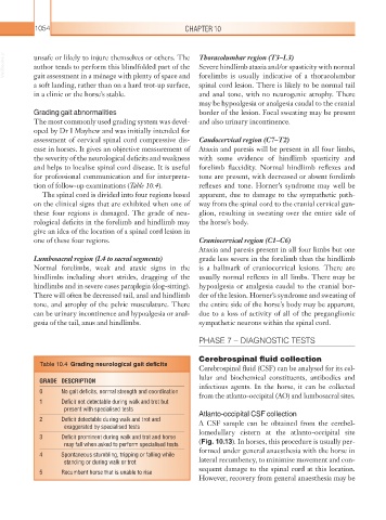
Table 10.4 Grading neurological gait deficits
Cerebrospinal fluid (CSF) can be analysed for its cel-
lular and biochemical constituents, antibodies and
GRADE DESCRIPTION
0 No gait deficits, normal strength and coordination infectious agents. In the horse, it can be collected
from the atlanto-occipital (AO) and lumbosacral sites.
1 Deficit not detectable during walk and trot but
present with specialised tests Atlanto-occipital CSF collection
2 Deficit detectable during walk and trot and A CSF sample can be obtained from the cerebel-
exaggerated by specialised tests
3 Deficit prominent during walk and trot and horse lomedullary cistern at the atlanto-occipital site
may fall when asked to perform specialised tests (Fig. 10.13). In horses, this procedure is usually per-
4 Spontaneous stumbling, tripping or falling while formed under general anaesthesia with the horse in
standing or during walk or trot lateral recumbency, to minimise movement and con-
5 Recumbent horse that is unable to rise sequent damage to the spinal cord at this location.
However, recovery from general anaesthesia may be