
Page 1257 - Equine Clinical Medicine, Surgery and Reproduction, 2nd Edition
P. 1257
1232 CHAPTER 12
VetBooks.ir NEOPLASTIC DISEASE cell degranulation with the release of many phar-
macologically active agents. The allergic response
Definition/overview
antigen-presenting cells and T lymphocytes, par-
Primary neoplasia in the horse may cause depres- has been further elaborated to involve Langerhans
sion and pain, but is rarely pruritic. Where pruri- ticularly T helper cells. The T helper-2 (Th2) cell,
tus occurs, examination for blow fly larvae or fungal in fact, produces cytokines such as interleukins
infection, such as pythiosis, is warranted. Habronema (IL)-4, -5, -6, -10 and -13, among which IL-4 and
spp. larvae have also been found in skin neoplasms IL-13 are essential for B cell immunoglobulin
on histopathological examination. class switching to IgE and IL-5 is key to recruit-
ment of eosinophils. In non-atopic individuals,
ATOPY the T helper-1 (Th1) cell line produces interferon
(IFN) and IL-2, which in turn suppress the pro-
Definition/overview liferation of allergy promoting Th2 cells, and are
Atopy is a genetically mediated pruritus and type 1 responsible for the local immune defence system.
hypersensitivity. T regulatory cells and IL-10 are now thought to
play a pivotal role in altering the balance between
Aetiology/pathophysiology Th1 and Th2 cells.
Breed predisposition in this disease tends to indicate
genetic programming. Predisposed breeds include Clinical presentation
Thoroughbreds, Quarter horses, Warmbloods, Recurrent, irregular, bilaterally symmetrical pruri-
Arabs and Morgans, with cases in males (usually tus, which may be related or unrelated to the sea-
geldings) almost twice as prevalent as cases in son of the year, is characteristic. This often occurs
mares, and a median age of onset between 5 and without any other clinical signs. Initially, there is
7 years (range 2–12 years). The classic description self-mutilation, with horses biting themselves and
is of immunoglobulin (Ig)E becoming tissue-fixed, rubbing raw areas on their body, limbs, head and/
especially to skin. Mast cell-fixed IgE then reacts or ears (Fig. 12.22). Secondary lesions are partly
with its specific allergen or allergens, causing mast related to self-inflicted damage, leading to excoria-
tion, alopecia, lichenification and hyperpigmenta-
tion. Some horses develop a sterile eosinophilic
folliculitis. Chronic recurrent urticaria and angi-
12.22 oedema, which may or may not be pruritic, and
allergen exacerbated recurrent inflammatory airway
disease, similar to asthma in humans and cats, may
either present singly or in combination with the
pruritic form. Some uncommon presenting signs
thought to be associated with allergies include lami-
nitis, otitis and head shaking.
Differential diagnosis
Hypersensitivity to food, pollens, moulds, dust and
many insects (ectoparasites).
Diagnosis
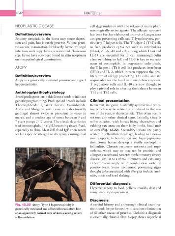
Fig. 12.22 Atopy. Type 1 hypersensitivity is A careful history and a thorough clinical examina-
genetically mediated and affected horses often bite tion should be performed, with absolute elimination
at an apparently normal area of skin, causing severe of all other causes of pruritus. Definitive diagnosis
self-mutilation. is essentially clinical. Skin biopsy shows superficial