
Page 225 - Equine Clinical Medicine, Surgery and Reproduction, 2nd Edition
P. 225
200 CHAPTER 1
VetBooks.ir medial femorotibial and femoropatellar joints and any OSTEOCHONDROSIS OF THE
FEMOROPATELLAR JOINT
inadvertent intra-articular injection of a counterirri-
tant could prove disastrous.
Management of a fractured patella is determined Definition/overview
by the fracture configuration. Horses with small The femoropatellar joint is one of the most com-
non- displaced, non-articular fractures can be treated mon sites for OCD in the horse, particularly in the
conservatively with box rest, and the prognosis for Warmblood and Thoroughbred. The disease usually
soundness is good. There are a limited number of presents in actively growing large individuals when
reports of treatment of medial patellar fractures in the foals or weanlings but may remain clinically silent
literature but those of the medial pole may be ame- until the animal starts active work. It may predispose
nable to arthroscopic removal. It has been postulated to the early onset of OA of this joint.
that up to 30% of the patella can be removed safely,
with smaller fragments having a good prognosis for Clinical presentation
future athletic function. However, it is the author’s The condition usually presents before 2 years of age
experience that larger fragments are harder to treat, and is particularly seen between 4 and 14 months
with patellar instability an issue particularly where the of age in excessively growing individuals. In some
attachment of the medial patellar ligament is com- animals the disease remains asymptomatic in their
pletely disrupted. Internal fixation is required for all early life but presents later either as their work pro-
other fracture configurations, although the extreme gramme increases between 3 and 6 years of age or
pull exerted on the patella by the quadriceps muscles where excessive wear and tear leads to OA of the joint.
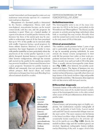
may promote breakdown. Comminuted fractures have There is usually obvious femoropatellar joint disten-
a poor prognosis. Treatment for patellar luxation is sion (Fig. 1.384), often bilateral, but possibly asym-
difficult and commonly does not provide significant metrical in degree. The degree of lameness can vary
athletic prospect. Both lateral release and medial from a mild bilateral stiffness of the hindlimb gait to a
imbrication techniques have been used. Breeding from marked unilateral lameness, especially where there are
affected animals should be avoided. large lesions of the lateral trochlear ridge and possible
lateral patella instability. The lameness often improves
with rest but returns with increased exercise.
1.384 Differential diagnosis
Traumatic lesions of the stifle joint and patella; sub-
chondral bone cysts of the medial femoral condyle;
sepsis of the femoropatellar joint; in the older animal
other causes of OA and inflammation of the femoro-
Fig. 1.384 patellar joint.
Extensive
synovial Diagnosis
effusion of the Clinical history and presenting signs are highly sus-
left hindlimb picious, but confirmation requires radiographs of
femoropatellar the stifle, preferably of both limbs. Lateral medial
joint in a and caudal 60° lateral craniomedial oblique views are
6-month-old the most useful. The most common site for lesions
Thoroughbred is the lateral trochlear ridge of the femur, although
with stifle occasionally lesions restricted to the articular sur-
osteochondrosis. face of the patella are seen (Fig. 1.385). Rarely, the
(Photo courtesy medial trochlear ridge and the trochlear groove are
Graham involved. Significant radiographic lesions include:
Munroe) marked flattening of the mid- to upper-third of the