
Page 633 - Equine Clinical Medicine, Surgery and Reproduction, 2nd Edition
P. 633
608 CHAPTER 3
VetBooks.ir 3.25 3.26
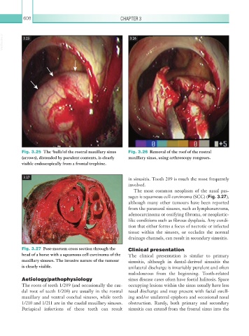
Fig. 3.25 The ‘bulla’of the rostral maxillary sinus Fig. 3.26 Removal of the roof of the rostral
(arrows), distended by purulent contents, is clearly maxillary sinus, using arthroscopy rongeurs.
visible endoscopically from a frontal trephine.
3.27 in sinusitis. Tooth 209 is much the most frequently
involved.
The most common neoplasm of the nasal pas-
sages is squamous cell carcinoma (SCC) (Fig. 3.27),
although many other tumours have been reported
from the paranasal sinuses, such as lymphosarcoma,
adenocarcinoma or ossifying fibroma, or neoplastic-
like conditions such as fibrous dysplasia. Any condi-
tion that either forms a focus of necrotic or infected
tissue within the sinuses, or occludes the normal
drainage channels, can result in secondary sinusitis.
Fig. 3.27 Post-mortem cross section through the Clinical presentation
head of a horse with a squamous cell carcinoma of the The clinical presentation is similar to primary
maxillary sinuses. The invasive nature of the tumour sinusitis, although in dental-derived sinusitis the
is clearly visible. unilateral discharge is invariably purulent and often
malodourous from the beginning. Tooth-related
Aetiology/pathophysiology sinus disease cases often have foetid halitosis. Space
The roots of teeth 1/209 (and occasionally the cau- occupying lesions within the sinus usually have less
dal root of teeth 1/208) are usually in the rostral nasal discharge and may present with facial swell-
maxillary and ventral conchal sinuses, while teeth ing and/or unilateral epiphora and occasional nasal
1/210 and 1/211 are in the caudal maxillary sinuses. obstruction. Rarely, both primary and secondary
Periapical infections of these teeth can result sinusitis can extend from the frontal sinus into the