
Page 737 - Equine Clinical Medicine, Surgery and Reproduction, 2nd Edition
P. 737
712 CHAPTER 3
VetBooks.ir 3.173 3.174
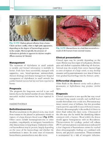
Fig. 3.173 Chylous pleural effusion from a horse.
Chyle can have a milky white to light pink appearance,
depending on the degree of haemorrhage present Fig. 3.174 Haemothorax in a foal that occurred as a
in the sample. Microscopically, the presence of result of rib fracture from external trauma.
chylomicron globules is apparent in chylous samples.
(Photo courtesy K Christie)
Clinical presentation
Clinical signs may be variable depending on the
Management cause. Horses may have signs of tachypnoea, distress,
The treatment of chylothorax in small animals pain and shallow respiration following rib fracture.
is variable and limited information is available in Animals may die acutely from severe haemorrhage
horses. Foals have been successfully managed with in cases of rupture of a large vessel, or may develop
supportive care, broad-spectrum antimicrobials, anaemia and hypoproteinaemia over time if there is
thoracic drainage and dietary management. Surgical more gradual haemorrhage into the thoracic cavity.
management of chylothorax in small animals has
yielded limited success but has not been reported in Differential diagnoses
horses. Other fluids in the thoracic cavity such as pleuro-
pneumonia or hydrothorax may produce similar
Prognosis signs.
The prognosis for long-term survival is not well
known due to the limited number of cases. However, Diagnosis
successful medical treatment has been reported in Clinical examination is non-specific but may reveal
foals. decreased lung sounds ventrally and muffled heart
sounds that radiate over a wide area. Percussion may
HAEMOTHORAX detect ventral areas of dullness, but this procedure
may be painful, especially in cases of thoracic trauma.
Definition/overview Thoracic ultrasonography is the method of choice to
Blood accumulation in the pleural cavity may result detect fluid within the chest cavity. Ultrasound of
from trauma to the pleural or pulmonary vessels or the ribs may also be useful for identifying defects
rupture of a large thoracic blood vessel (Fig. 3.174). consistent with a fracture. Blood within the thorax
Other causes include haemangiosarcoma or other should appear homogeneous with no flocculation.
neoplasia, coagulopathy or iatrogenic haemothorax Thoracocentesis for cytology, packed cell volume
after thoracotomy or lung biopsy. Unilateral or bilat- (PCV) and total protein concentration is indicated if
eral haemothorax may be present, largely depending a clear diagnosis of haemothorax cannot be reached
on the cause of the haemorrhage and whether the when ultrasonographic findings are combined with
mediastinum is intact. In some cases of traumatic history and physical examination. Thoracocentesis
haemothorax, pneumothorax may also occur. is accompanied by a risk of iatrogenic infection.