
Page 825 - Equine Clinical Medicine, Surgery and Reproduction, 2nd Edition
P. 825
800 CHAPTER 4
VetBooks.ir 4.124 4.125
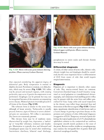
Fig. 4.125 Horse with acute grass sickness showing
bilateral upper eyelid ptosis. (Photo courtesy
Graham Munroe)
paraphimosis in entire males and chronic rhinitis
sicca may be noted.
Differential diagnosis
Fig. 4.124 Horse with acute grass sickness showing A variety of other causes of acute colic, chronic colic,
ptyalism. (Photo courtesy Graham Munroe) chronic weight loss and dysphagia should be consid-
ered, but the most important factor is differentiation
of EGS from causes of colic that would require
surgical intervention.
than expected considering the apparent degree of
abdominal pain. Body temperature is normal to Diagnosis
slightly elevated. Pytalism is common, as is dehydra- Palpation p/r is important to identify other causes
tion, which may be severe (Fig. 4.124). NG reflux of colic. Dry, mucus-covered faeces are common.
may be present and stomach rupture, with ensuing Concurrent distended loops of small intestine recog-
peritonitis, may occur if gastric decompression is not nised on rectal palpation or transabdominal ultraso-
performed. Dysphagia can be present but may not nography, and voluminous foul-smelling gastric reflux
be recognised because of the anorexia accompanying may be present with the acute form and reflect gen-
severe disease. Bilateral ptosis is invariably present in eralised GI ileus. Large-colon and caecal impactions
all forms of the disease (Fig. 4.125). in the chronic cases reflect large intestinal ileus and
The subacute form is similar to the acute form, desiccation of the fibrous ingesta (Fig. 4.126). Clinical
although of lesser severity. Tachycardia is usually signs, signalment, identification of risk factors such as
present; however, signs of distress or severe abdomi- a recent change in grazing or previous disease on the
nal pain are uncommon. NG reflux is uncommon. premises and exclusion of other differential diagnoses
Dry faeces are commonly present. is suggestive of EGS. Temporary reversal of ptosis fol-
The chronic form may be of insidious onset, lowing the topical application of a 0.5% phenylephrine
characterised by weight loss, depression, a ‘tucked- ophthalmic solution to the conjunctival sac confirms
up’ or ‘wasp-like’ abdomen, weakness and dyspha- neurogenic smooth muscle paralysis as the cause of
gia. Mastication is typically slow and laboured and the ptosis. Barium swallow studies or oesophagoscopy
oesophageal spasm may be noted after swallowing. may confirm the retrograde flow of gastric fluid and
Intermittent diarrhoea, bilateral nasal discharge, abnormal oesophageal motility.