
Page 376 - Fluid, Electrolyte, and Acid-Base Disorders in Small Animal Practice
P. 376
366 FLUID THERAPY
compared with forcing the catheter through unbroken outer surface of the catheter. Therefore the catheter
skin. The catheter should be tunneled subcutaneously should be removed as soon as possible.
as far parallel as practical before it enters the vein. As long
as the wound is no larger than the catheter diameter, the EMERGENCY CUT-DOWN
dermis will form a tight seal around the catheter shaft to PROCEDURE
limit bacterial migration from the skin. An emergency cut down is used to cannulate a vein when
attempts at percutaneous catheterization have failed or
MINICUT-DOWN PROCEDURE are likely to fail in a patient that requires immediate
This approach is the same as the facilitation procedure, venous access. This is an essential skill for emergency
but the incision is sufficiently extended so that the vessel’s clinicians that should be considered for any patient
sides and superficial surface are visible. The vessel may requiring immediate venous access. Any vein may be
then be catheterized under direct visualization, or it is used, but the author prefers the lateral saphenous vein
carefully dissected free of surrounding tissue, elevated in dogs (Figure 15-6, A) because the thin skin overlying
from the wound, incised with the bevel of a 20-gauge this vein facilitates access, and the vein may be successfully
needle, and then catheterized. This procedure is best and rapidly isolated with shaking hands. With practice,
done on any superficial vessel that has not been previously you should be able to catheterize this vein within 30 to
traumatized by percutaneous attempts. It is a reliable 60 seconds.
technique when direct percutaneous catheterization is 1. If time permits, clip the hair and cleanse the skin. This
difficult because of vascular collapse. However, the resul- step may be omitted in patients with short hair-coats
tant skin wound promotes bacterial migration along the that require immediate access; if the hair coat is long
A
B C
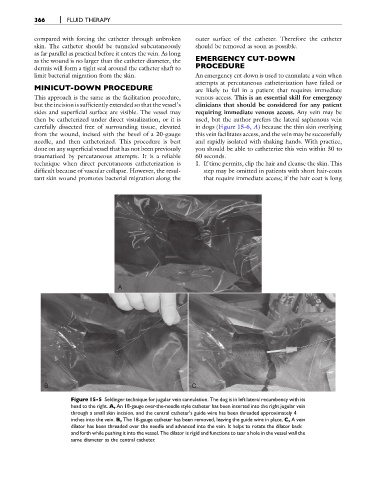
Figure 15-5 Seldinger technique for jugular vein cannulation. The dog is in left lateral recumbency with its
head to the right. A, An 18-gauge over-the-needle style catheter has been inserted into the right jugular vein
through a small skin incision, and the central catheter's guide wire has been threaded approximately 4
inches into the vein. B, The 18-gauge catheter has been removed, leaving the guide wire in place. C, A vein
dilator has been threaded over the needle and advanced into the vein. It helps to rotate the dilator back
and forth while pushing it into the vessel. The dilator is rigid and functions to tear a hole in the vessel wall the
same diameter as the central catheter.