
Page 650 - Fluid, Electrolyte, and Acid-Base Disorders in Small Animal Practice
P. 650
Enteral Nutrition 637
The placement of a NJ tube (Figure 26-13) involves a
minimally invasive technique that can be done under
intravenous sedation or a short anesthetic period
(Figures 26-14 and 26-15). Techniques of nasojejunal
tube intubation in dogs have recently been
described. 6,7,80,100 With experience these tubes can be
placed in less than 15 minutes. The weighted tip is coated
with lidocaine gel as a local anesthetic. A suture is placed
around the feeding tube with a purse-string and Chinese
finger trap pattern using a 2-0 to 3-0 synthetic or silk
suture material. Nasojejunal feeding tubes should be
secured with nonabsorbable suture at the nostril and side
of the cheek. NJ feeding tubes are best suited for short-
term (<1 week) delivery of postpyloric enteral nutritional
B
in hospitalized patients. Administration of nutrients to
the jejunum has minimal effect on pancreatic secretion
and feedings can be continued despite vomiting. 81,84
A
Figure 26-12 Percutaneous nonendoscopic gastrostomy tube
placement. Passage of the placement device with animal in right
lateral recumbency. Palpation of the device in the stomach is
facilitated by positioning the animal's head over the end of the table
and extended: A, Tube placement device rotated 90 degrees
counterclockwise to pass freely over the base of the heart. B, The
catheter needle is inserted through the skin into the flared end of the
device at a 45-degree angle parallel with the distal end of the device.
(Drawing by Tim Vojt).
BOX 26-4 List of Materials
Needed for
Percutaneous
Gastrostomy Tube
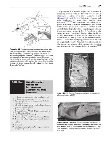
Placement Figure 26-13 Common feeding tubes adapted for nasojejunal
placement in dogs at MSU.
1. Endoscope or gastrostomy tube introduction set
2. Endoscope grasping forceps
3. Bard urologic catheter or polyurethane PEG with
collapsible bumper
4. 14- to 22-gauge peripheral or indwelling catheter, or D
open end tom-cat catheter
5. Three-way stopcock J
6. Braunamid suture (Vetafil) 2-0; approximately 2 ft A
7. No. 11 surgical blade
8. 18- to 20-gauge, 1-inch needles
9. Rubber tubing (1.5 inch long) for external flange
10. 1-inch wide adhesive tape; approximately 6 inches
long
11. Scissors Figure 26-14 Right lateral thoraco-abdominal radiograph of a
12. Hemostats recumbent dog showing the correct positioning of the nasojejunal
13. Bandage material optional; Vet wrap, stockinette, tube with the distal tip in the jejunum just before removal (5 days
cast padding, or Kling after placement). D, descending duodenum; A, ascending duodenum;
J, jejunum.