
Page 1069 - Adams and Stashak's Lameness in Horses, 7th Edition
P. 1069
Lameness in the Young Horse 1035
the physis. Directly under this perichondrial ring is the Recently it has been shown that this zone contains cells
45
zone of Ranvier at the level of the resting and proliferative that can act as precursors of the cells in the proliferative
VetBooks.ir undifferentiated cells, fibroblasts, and progenitor cells that the cells line up into parallel columns along the long
zone and it produces an orienting factor that dictates
zones (Figure 10.4). This wedge‐shaped zone contains
34
axis of the bone.
for osteoblasts that are supplied to the resting/reserve
1
52
zone. These cells function to expand the diameter of
the growth plate. 52
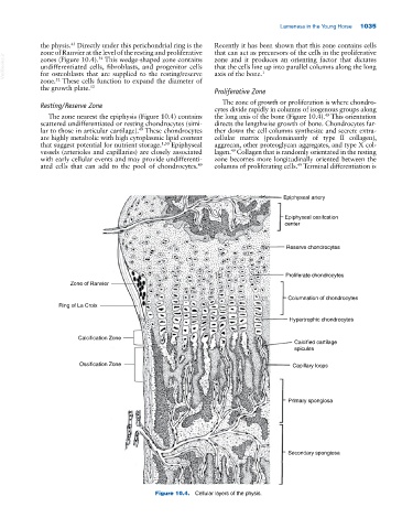
Proliferative Zone
The zone of growth or proliferation is where chondro
Resting/Reserve Zone
cytes divide rapidly in columns of isogenous groups along
The zone nearest the epiphysis (Figure 10.4) contains the long axis of the bone (Figure 10.4). This orientation
49
scattered undifferentiated or resting chondrocytes (simi directs the lengthwise growth of bone. Chondrocytes far
lar to those in articular cartilage). These chondrocytes ther down the cell columns synthesize and secrete extra
49
are highly metabolic with high cytoplasmic lipid content cellular matrix (predominantly of type II collagen),
that suggest potential for nutrient storage. 1,50 Epiphyseal aggrecan, other proteoglycan aggregates, and type X col
vessels (arterioles and capillaries) are closely associated lagen. Collagen that is randomly orientated in the resting
49
with early cellular events and may provide undifferenti zone becomes more longitudinally oriented between the
49
ated cells that can add to the pool of chondrocytes. columns of proliferating cells. Terminal differentiation is
49
Epiphyseal artery
Epiphyseal ossifcation
center
Reserve chondrocytes
Proliferate chondrocytes
Zone of Ranvier
Columnation of chondrocytes
Ring of La Croix
Hypertrophic chondrocytes
Calcification Zone
Calcified cartilage
spicules
Ossification Zone Capillary loops
Primary spongiosa
Secondary spongiosa
Figure 10.4. Cellular layers of the physis.