
Page 1130 - Adams and Stashak's Lameness in Horses, 7th Edition
P. 1130
1096 Chapter 11
VetBooks.ir
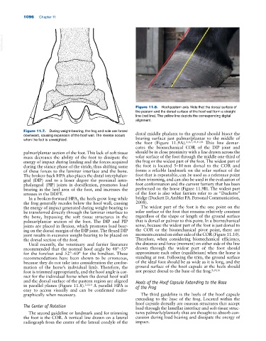
Figure 11.8. Hoof‐pastern axis. Note that the dorsal surface of
the pastern and the dorsal surface of the hoof wall form a straight
line (red line). The yellow line depicts the corresponding digital
alignment.
Figure 11.7. During weight‐bearing, the frog and sole are forced distal middle phalanx to the ground should bisect the
downward, causing expansion of the hoof wall. The reverse occurs bearing surface just palmar/plantar to the middle of
when the foot is unweighted.
the foot (Figure 11.9A). 4,6,7,10,11,14 This line demar
cates the biomechanical COR of the DIP joint and
palmar/plantar section of the foot. This lack of soft tissue should be in close proximity with a line drawn across the
mass decreases the ability of the foot to dissipate the solar surface of the foot through the middle one‐third of
energy of impact during landing and the forces acquired the frog or the widest part of the foot. The widest part of
during the stance phase of the stride, thus shifting some the foot is located 5–10 mm dorsal to the COR and
of these forces to the laminar interface and the bone. forms a reliable landmark on the solar surface of the
The broken‐back HPA also places the distal interphalan foot that is repeatable, can be used as a reference point
geal (DIP) and to a lesser degree the proximal inter when trimming, and can also be used in the evaluation of
phalangeal (PIP) joints in dorsiflexion, promotes load foot conformation and the current farriery that has been
bearing in the heel area of the foot, and increases the performed on the horse (Figure 11.9B). The widest part
stresses in the DDFT. of the foot is also what farriers refer to as “Ducketts”
In a broken‐forward HPA, the heels grow long while bridge (Duckett D, Ambler PA. Personal Communication,
the frog generally recedes below the hoof wall, causing 2008).
the energy of impact generated during weight‐bearing to The widest part of the foot is the one point on the
be transferred directly through the laminar interface to solar surface of the foot that remains relatively constant
the bone, bypassing the soft tissue structures in the regardless of the shape or length of the ground surface
palmar/plantar section of the foot. The DIP and PIP that is dorsal or palmar to this point. In a biomechanical
joints are placed in flexion, which promotes load bear sense, because the widest part of the foot is just dorsal to
ing on the dorsal margin of the DIP joint. The flexed DIP the COR or the biomechanical pivot point, there are
joint results in excessive weight‐bearing to be placed on moments created on either side of the COR (Figure 11.10).
the dorsal section of the foot. Therefore, when considering biomechanical efficiency,
Until recently, the veterinary and farrier literature the distance and force (moment) on either side of the line
recommended that the normal hoof angle be 48°–55° drawn through the widest part of the foot should
for the forefeet and 52°–60° for the hindfeet. These approximate each other (equilibrium) when the horse is
recommendations have been shown to be erroneous, standing at rest. Following the trim, the ground surface
because they do not take into consideration the confor of the ideal foot should be as wide as it is long, and the
mation of the horse’s individual limb. Therefore, the ground surface of the hoof capsule at the heels should
foot is trimmed appropriately, and the hoof angle is cor not project dorsal to the base of the frog. 8,14,16
rect for the individual horse when the dorsal hoof wall
and the dorsal surface of the pastern region are aligned Heels of the Hoof Capsule Extending to the Base
in parallel planes (Figure 11.8). 2,3,14 A parallel HPA is of the Frog
easy to access visually and can be confirmed radio
graphically when necessary. The third guideline is the heels of the hoof capsule
extending to the base of the frog. Located within the
hoof capsule dorsally are osseous structures that accept
The Center of Rotation load through the lamellae interface and soft tissue struc
The second guideline or landmark used for trimming tures palmarly/plantarly that are thought to absorb con
the foot is the COR. A vertical line drawn on a lateral cussion during load bearing and dissipate the energy of
radiograph from the center of the lateral condyle of the impact.