
Page 1213 - Adams and Stashak's Lameness in Horses, 7th Edition
P. 1213
Miscellaneous Musculoskeletal Conditions 1179
a ballpoint pen. Mild stimulation of the chest and abdo
men elicits a cutaneous trunci muscle response, but not
VetBooks.ir to elicit this local reflex first, and then a pain response
in all horses. A two‐pinch technique with forceps is used
may be noticed with a deeper pinch.
The dorsal processes of the thoracic and lumbar ver
tebrae must be palpated and (gently) percussed. No pain
response or crepitus should be observed or palpated.
With both hands placed over the withers, the horse is
pushed away and pulled toward the examiner. A normal
horse quickly activates the extensor or antigravity mus
cles in the forelimbs to maintain a solid stance. An
abnormal horse can easily be pushed away or pulled
toward the examiner because of LMN weakness or a
lack of proprioception.
Hopping can be done by picking up one forelimb at a
time; however, caution is advised in the obviously
severely affected horse. The examiner faces the same
direction as the horse, holds the lifted forelimb at the
pasterns, and pushes the horse away with his/her shoul
der. The normal horse will hop one jump at a time with
a nice lift‐off and landing. The abnormal horse will have
a delayed lift‐off and a short, rigid jump followed by a
clumsy landing. “Hopping” allows to test several aspects
of the nervous system: muscular strength, propriocep
tion, and dysmetria.
Stimulating the axial musculature of the back usually
elicits a ventrodorsal movement of the vertebral column,
lordosis followed by kyphosis. The abnormal horse
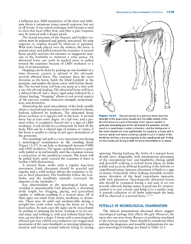
shows scoliosis or it appears stiff in the back. A normal Figure 12.27. Tail pull (arrow) in a standing horse tests the
horse has at least some degree of a tail tone and a per strength of the quadriceps muscle and the reflex activity of the
ineal reflex. A completely flaccid tail is a sign of LMN femoral nerve as a part of the lower motor neuron system. A
weakness. A tail should not continuously be held off the gradually increasing pull on the tail toward the examiner should
body. This can be a clinical sign of tetanus or tetany, if result in a quadriceps muscle contraction, and the hindquarter of
the horse is unable to clamp its tail upon stimulation of the horse should not move significantly. For example, a horse with a
the perineum. cervical spinal cord lesion showing a grade 3 out of 4 ataxia of the
The (standing) tail pull is a very important test during hindlimbs still has a strong response to the standing tail pull. Pulling
a neurological examination in the standing horse on this horse’s tail during a walk will show incoordination or ataxia.
(Figure 12.27). It can help to distinguish between UMN
and LMN weakness. The square standing horse is grad
ually pulled at its tail laterally until the examiner notices spinning. During backing, the limbs of a normal horse
a contraction of the quadriceps muscle. The horse will should move diagonally, with simultaneous placement
be pulled fairly easily toward the examiner if there is of the contralateral fore‐ and hindlimbs. During uphill
lumbar LMN dysfunction. and downhill walking, a normal horse places its limbs
A normal horse walks with a regular four‐beat solidly and securely, without knuckling in the fetlocks or
movement. Limb placement should be graceful and carpal/tarsal joints. Insecure placement of limbs is a sign
regular, and a solid surface allows the examiner to lis of ataxia. Particularly when walking downhill, simulta
ten to hoof placement. The hindlimbs follow the fore neous elevation of the head exacerbates insecurity
limbs, and the hindlimbs typically overreach the with limb placement. Neurologically abnormal horses
imprints of the forelimbs. 10 also should be examined during a trot and, if not too
Key abnormalities in the neurological horse are severely affected, during canter. A good test for proprio
revealed in unpredictable limb placement, a shortened ception is to trot a horse and bring it to a sudden stop.
stride length, toe dragging, pacing, and generalized A smooth collection of all limbs underneath the body
rigidity or stiffness resulting in a truncal sway. All of will be observed in a normal horse. 1,2,5,10
these signs can result from ataxia, weakness, or dysme
tria. These may be mild and unobservable during a
straight‐line exam when walking the horse on a flat PITFALLS OF NEUROLOGICAL EXAMINATION
hard surface. In such cases the signs can be exacerbated
by circling or spinning the horse, backing it up for sev The clinical presentations discussed above suggest
eral steps, and walking it, with and without head eleva neurological damage that affects the gait. However, the
tion, up and down a slope. A horse with a neurologically signs also can arise from illnesses or problems unrelated
affected gait may exhibit circumduction (an exaggerated to the nervous system. These should be considered when
movement of the outer hindlimb) or pivoting (planting a making the diagnosis, and possible explanations for sus
forefoot and turning around without lifting it) during pect neurological findings are listed in Table 12.5.