
Page 143 - Adams and Stashak's Lameness in Horses, 7th Edition
P. 143
Examination for Lameness 109
The semimembranosus and semitendinosus muscles rupture of the peroneus tertius muscle are present dur
should be palpated for any evidence of pain and swell ing exercise. With the stifle flexed, the hock can be
VetBooks.ir scarring/fibrosis that is often present with fibrotic myo nemius tendon occurs (Figure 2.89).
extended, and a characteristic dimpling of the gastroc
ing suggestive of myositis (hamstring pull) and for firm
pathy (Figure 2.88). Although an uncommon site for
problems, the gastrocnemius tendon should be palpated
for swelling and pain. An attempt should also be made STIFLE
to extend the hock joint if clinical signs consistent with
The stifle should be observed and palpated for swelling
and/or atrophy of the associated muscle groups and for
fluid distension of the joints. Distension of the femoropatellar
joint is best seen from the lateral view (Figure 2.90),
and distension of the medial femorotibial (MFT) is best
observed from the cranial aspect (Figure 2.91). However,
palpation is the preferred method to detect effusion within
the stifle joints. The femoropatellar joint pouch is located
on the cranial aspect of the stifle beneath the patella liga
ments. In general, effusion of the femoropatellar joint
makes palpation of the three distal patellar ligaments
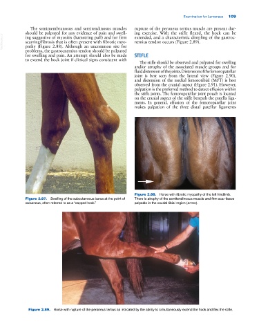
Figure 2.88. Horse with fibrotic myopathy of the left hindlimb.
Figure 2.87. Swelling of the subcutaneous bursa at the point of There is atrophy of the semitendinosus muscle and firm scar tissue
calcaneus, often referred to as a “capped hock.” palpable in the caudal tibial region (arrow).
Figure 2.89. Horse with rupture of the peroneus tertius as indicated by the ability to simultaneously extend the hock and flex the stifle.