
Page 39 - Adams and Stashak's Lameness in Horses, 7th Edition
P. 39
Functional Anatomy of the Equine Musculoskeletal System 5
VetBooks.ir
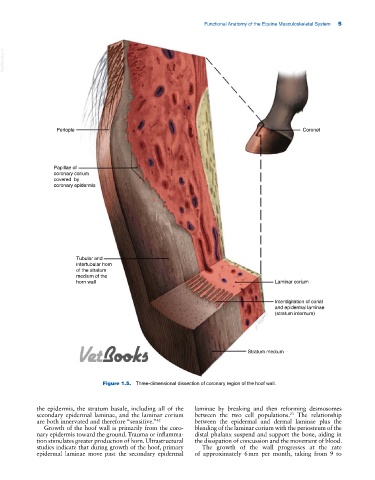
Periople Coronet
Papillae of
coronary corium
covered by
coronary epidermis
Tubular and
intertubular horn
of the stratum
medium of the
horn wall Laminar corium
Interdigitation of corial
and epidermal laminae
(stratum internum)
Stratum medium
Figure 1.5. Three‐dimensional dissection of coronary region of the hoof wall.
the epidermis, the stratum basale, including all of the laminae by breaking and then reforming desmosomes
secondary epidermal laminae, and the laminar corium between the two cell populations. The relationship
23
are both innervated and therefore “sensitive.” 42 between the epidermal and dermal laminae plus the
Growth of the hoof wall is primarily from the coro blending of the laminar corium with the periosteum of the
nary epidermis toward the ground. Trauma or inflamma distal phalanx suspend and support the bone, aiding in
tion stimulates greater production of horn. Ultrastructural the dissipation of concussion and the movement of blood.
studies indicate that during growth of the hoof, primary The growth of the wall progresses at the rate
epidermal laminae move past the secondary epidermal of approximately 6 mm per month, taking from 9 to