
Page 459 - Adams and Stashak's Lameness in Horses, 7th Edition
P. 459
Diagnostic Imaging 425
VetBooks.ir
A B
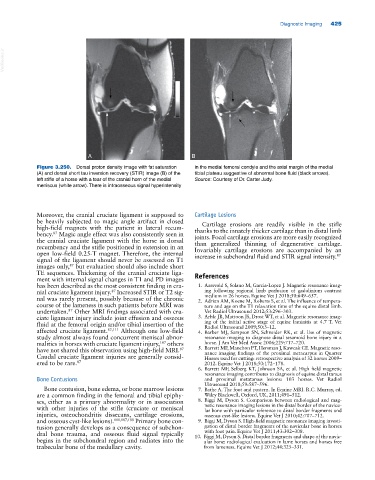
Figure 3.250. Dorsal proton density image with fat saturation in the medial femoral condyle and the axial margin of the medial
(A) and dorsal short tau inversion recovery (STIR) image (B) of the tibial plateau suggestive of abnormal bone fluid (black arrows).
left stifle of a horse with a tear of the cranial horn of the medial Source: Courtesy of Dr. Carter Judy.
meniscus (white arrow). There is intraosseous signal hyperintensity
Moreover, the cranial cruciate ligament is supposed to Cartilage Lesions
be heavily subjected to magic angle artifact in closed Cartilage erosions are readily visible in the stifle
high‐field magnets with the patient in lateral recum thanks to the innately thicker cartilage than in distal limb
87
bency. Magic angle effect was also consistently seen in joints. Focal cartilage erosions are more easily recognized
the cranial cruciate ligament with the horse in dorsal than generalized thinning of degenerative cartilage.
recumbency and the stifle positioned in extension in an Invariably cartilage erosions are accompanied by an
open low‐field 0.25‐T magnet. Therefore, the internal increase in subchondral fluid and STIR signal intensity. 87
signal of the ligament should never be assessed on T1
images only, but evaluation should also include short
87
TE sequences. Thickening of the cranial cruciate liga References
ment with internal signal changes in T1 and PD images
has been described as the most consistent finding in cra 1. Aarsvold S, Solano M, Garcia‐Lopez J. Magnetic resonance imag
nial cruciate ligament injury. Increased STIR or T2 sig ing following regional limb perfusion of gadolinium contrast
87
medium in 26 horses. Equine Vet J 2018;50:649–657.
nal was rarely present, possibly because of the chronic 2. Adrian AM, Koene M, Roberts S, et al. The influence of tempera
course of the lameness in such patients before MRI was ture and age on the T1 relaxation time of the equine distal limb.
undertaken. Other MRI findings associated with cru Vet Radiol Ultrasound 2012;53:296–303.
87
ciate ligament injury include joint effusion and osseous 3. Arble JB, Mattoon JS, Drost WT, et al. Magnetic resonance imag
fluid at the femoral origin and/or tibial insertion of the ing of the initial active stage of equine laminitis at 4.7 T. Vet
Radiol Ultrasound 2009;50:3–12.
affected cruciate ligament. 87,111 Although one low‐field 4. Barber MJ, Sampson SN, Schneider RK, et al. Use of magnetic
study almost always found concurrent meniscal abnor resonance imaging to diagnose distal sesamoid bone injury in a
malities in horses with cruciate ligament injury, others horse. J Am Vet Med Assoc 2006;229:717–720.
187
have not shared this observation using high‐field MRI. 5. Barrett MF, Manchon PT, Hersman J, Kawcak CE. Magnetic reso
87
nance imaging findings of the proximal metacarpus in Quarter
Caudal cruciate ligament injuries are generally consid Horses used for cutting: retrospective analysis of 32 horses 2009–
ered to be rare. 87 2012. Equine Vet J 2018;50:172–178.
6. Barrett MF, Selberg KT, Johnson SA, et al. High field magnetic
resonance imaging contributes to diagnosis of equine distal tarsus
Bone Contusions and proximal metatarsus lesions: 103 horses. Vet Radiol
Ultrasound 2018;59:587–596.
Bone contusion, bone edema, or bone marrow lesions 7. Bathe A. The foot and pastern. In Equine MRI. R.C. Murray, ed.
are a common finding in the femoral and tibial epiphy Wiley Blackwell, Oxford, UK, 2011;491–512.
ses, either as a primary abnormality or in association 8. Biggi M, Dyson S. Comparison between radiological and mag
with other injuries of the stifle (cruciate or meniscal netic resonance imaging lesions in the distal border of the navicu
lar bone with particular reference to distal border fragments and
injuries, osteochondritis dissecans, cartilage erosions, osseous cyst‐like lesions. Equine Vet J 2010;42:707–712.
and osseous cyst‐like lesions). 111,187,188 Primary bone con 9. Biggi M, Dyson S. High‐field magnetic resonance imaging investi
tusion generally develops as a consequence of subchon gation of distal border fragments of the navicular bone in horses
with foot pain. Equine Vet J 2011;43:302–308.
dral bone trauma, and osseous fluid signal typically 10. Biggi M, Dyson S. Distal border fragments and shape of the navic
begins in the subchondral region and radiates into the ular bone: radiological evaluation in lame horses and horses free
trabecular bone of the medullary cavity. from lameness. Equine Vet J 2012;44:325–331.