
Page 466 - Adams and Stashak's Lameness in Horses, 7th Edition
P. 466
432 Chapter 3
A 96.0 °F B convection are responsible for heat loss below that tem
perature. Very cold environmental temperatures may
VetBooks.ir 95 cause vasoconstriction of the lower legs and interfere
with imaging. In these cases, low‐level exercise to stim
ulate vasodilatation is necessary. The thermographic
area ideally should have a steady, uniform airflow so
that erroneous cooling does not occur. Practically, the
90 horse should be kept from drafts. Likewise, the horse
should be allowed 10–20 minutes to acclimate to the
environment or room where thermography is per
formed. Artifacts are extraneous sources on the skin
C 85 D that can cause irregular images. Among these are debris,
scar tissue, hair length, liniments, leg wraps, and equip
ment. To avoid artifacts, make sure all subjects are
21
groomed and free of leg wraps and equipment for 2
hours whenever possible. Hair insulates the leg and
80
blocks the emission of infrared radiation. But, as long
as the hair is short and of uniform length, the thermal
image produced is accurate. The skin should always be
76.0 evaluated for changes in hair length that may cause
false “hot spots” in the thermogram.
Multiple thermographic images of a suspect area
should be made. 25,26 The area in question should be eval
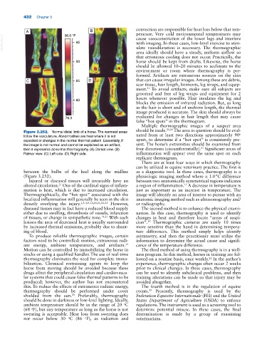
Figure 3.252. Normal distal limb of a horse. The warmest areas o
follow the vasculature. Abnormalities are heat where it is not uated from at least two directions approximately 90
expected or changes in the normal thermal pattern. Essentially, if apart, to determine if a “hot spot” is consistently pre
the image is not normal and cannot be explained as an artifact, sent. The horse’s extremities should be examined from
21
then it represents abnormal thermography. (A) Dorsal view. (B) four directions (circumferentially). Significant areas of
Palmar view. (C) Left side. (D) Right side. inflammation will appear over the same spot on each
replicate thermogram.
There are at least four ways in which thermography
can be utilized in equine veterinary practice. The first is
between the bulbs of the heel along the midline as a diagnostic tool. In these cases, thermography is a
(Figure 3.252). physiologic imaging method where a 1.0 °C difference
Injured or diseased tissues will invariably have an between two anatomically symmetrical regions indicates
altered circulation. One of the cardinal signs of inflam a region of inflammation. A decrease in temperature is
21
15
mation is heat, which is due to increased circulation. just as important as an increase in temperature. The
Thermographically, the “hot spot” associated with the image will identify an area of interest to pursue with an
localized inflammation will generally be seen in the skin anatomic imaging method such as ultrasonography and/
directly overlying the injury. 1,3–5,9–14,20,21,23–25 However, or radiography.
diseased tissues may in fact have a reduced blood supply The second method is to enhance the physical exami
either due to swelling, thrombosis of vessels, infarction nation. In this case, thermography is used to identify
of tissues, or change in sympathetic tone. 14,21 With such changes in heat and therefore locate “areas of suspi
lesions the area of decreased heat is usually surrounded cion”. Thermographic cameras are easily 10 times
15
by increased thermal emissions, probably due to shunt more sensitive than the hand in determining tempera
ing of blood. ture differences. This method simply helps identify
To produce reliable thermographic images, certain asymmetry, and then the practitioner must utilize the
factors need to be controlled: motion, extraneous radi information to determine the actual cause and signifi
ant energy, ambient temperature, and artifacts. cance of the temperature difference.
15
Motion can be controlled by immobilizing the horse in The third method of using thermography is in a well
stocks or using a qualified handler. The use of real‐time ness program. In this method, horses in training are fol
thermography eliminates the need for complete immo lowed on a routine basis, once weekly. In the author’s
22
bilization. Chemical restraining agents to keep the experience, thermographic changes often occur 2 weeks
horse from moving should be avoided because these prior to clinical changes. In these cases, thermography
drugs affect the peripheral circulation and cardiovascu can be used to identify subclinical problems, and then
lar systems that could cause false thermal patterns to be training alterations can be made so that injury may be
produced; however, the author has not encountered avoided altogether.
this. To reduce the effects of extraneous radiant energy, The fourth method is in the regulation of equine
thermography should be performed under cover events. Presently, thermography is used by the
15
21
shielded from the sun. Preferably, thermography Federation Equestre Internationale (FEI) and the United
should be done in darkness or low‐level lighting. Ideally, States Department of Agriculture (USDA) to enforce
ambient temperature should be in the range of 20 C regulations. The instrument is used as a screening tool to
o
(68 F), but any temperature as long as the horse is not determine potential misuse. In these cases, the final
o
sweating is acceptable. Heat loss from sweating does determination is made by a group of examining
not occur below 30 C (86 F), as radiation and veterinarians.
o
o